
Applications
 Part of the Oxford Instruments Group
Expand
Collapse
Part of the Oxford Instruments Group

Application Notes
Author: Ute Schmidt, Eleni Vomhof
Published: 16 Oct 2026 · Last updated: 16 Oct 2026
Characterising 2D materials like graphene, transition metal dichalcogenides (TMDs), and hexagonal boron nitride (hBN) typically requires combining multiple optical techniques because each method reveals different physical properties. Without an automated workflow, the user must manually switch between techniques and initiate each measurement.
This article presents a workflow that integrates Raman, photoluminescence (PL), and polarisation-resolved second harmonic generation (SHG) imaging into one automated measurement routine. As all measurements are performed at the same sample position, the resulting image set enables direct comparison of the complementary information provided by the three techniques:
The automated workflow is particularly relevant for high-throughput characterisation, core facilities, and industrial environments that require reproducible multimodal measurements.
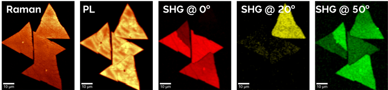
Fig. 1: Representative image examples of WS2 flakes obtained with the automated workflow. From left to right: Raman image, PL image, and SHG images recorded at 0°, 20°, and 50° polarisation angle of the incoming laser wave.
Compared to manual workflows, the automated approach reduces user intervention allowing for high throughput and reproducibility in routine measurements (Table 1). Automation enables:
Table 1: Comparison of manual and automated workflow.
| Feature | Manual Approach | Automated Workflow |
| Setup per technique | Required | Single setup |
| Alignment | Repeated | Fixed and consistent |
| Measurement | User intervention | No user intervention |
| Throughput | Low | High |
| Reproducibility | Variable results | Assured consistent results |
The workflow consists of a one-time microscope setup (Fig. 2, bright blue steps) followed by a fully automated acquisition sequence (Fig. 2, dark blue steps). Instead of starting each scan manually, the user defines the scan area. Then the system executes all measurement modalities point by point and line by line and stores co-registered datasets for direct comparison.
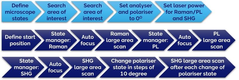
Fig. 2: Workflow for correlative Raman, PL and SHG imaging of 2D materials. Bright blue steps: Setting up the witec360 microscope. Dark blue steps: Automated workflow.
The automated workflow and exemplary measurements presented in this article rely on a fully automated witec360 microscope with the following equipment configuration:
The acquisition sequence (dark blue steps of the workflow in Fig. 2) was automated using the Workflow Manager of the WITec Suite software.
Tungsten disulfide (WS2) is a typical 2D semiconductor material used in optoelectronics and valleytronics. WS2 flakes on Si/SiO2 substrates were characterised using the automated, multimodal microscopy workflow integrating Raman, PL and polarised SHG imaging.
Fig. 3 shows the bright-field image of the area of interest. A common scan area was defined for Raman, PL and SHG imaging, indicated by the red box in Fig. 3. All measurements were performed sequentially by the automated routine without user intervention. High-resolution maps were acquired by raster scanning the sample with a fixed step size to ensure dense spatial sampling. At each position, Raman spectra, PL emission, and SHG signals were recorded under their respective excitation conditions while maintaining consistent spatial alignment. SHG images were acquired at seven distinct incident-polarisation angles revealing the crystallographic orientation of the WS2 crystals. The parameters for the automated scans are set in the Large Area Scan in the Control Window of the WITec Control software and listed in Table 2.
The resulting datasets are intrinsically co-registered in the WITec Suite software and analysed to correlate structural, optical, and symmetry-related properties across the sample.
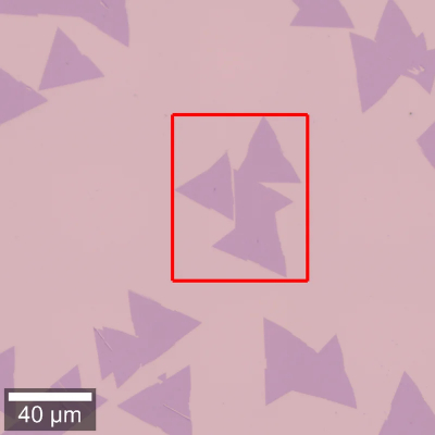
Fig. 3: Bright-field image of WS2 flakes, with the red box highlighting the area of interest for further investigation by Raman, PL and SHG.
Table 2: Imaging parameters common to all measurements performed with this workflow.
| Imaging Parameter | Objective | Area | Pixels | Integration time per pixel |
| 100x/0.9 | 65 x 80 µm² | 130x160 | 0.05 s |
Figure 4 shows the nine co-registered Raman, PL and SHG datasets acquired automatically from the same sample region.
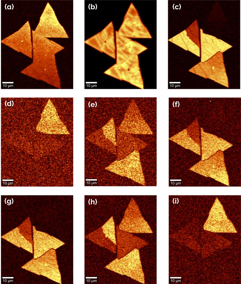
Fig. 4: Raman, PL and SHG image set of WS2 flakes acquired using the automated measurement workflow. (a) Raman image, (b) PL image, and SHG images recorded at incident laser polarisation angles of (c) 0°, (d) 10°, (e) 20°, (f) 30°, (g) 40°, (h) 50°, and (i) 60°. All images were generated using SUM filtering over the respective spectral peaks.
The complementary information from the three techniques enables comprehensive characterisation of the WS2 sample.
Raman imaging: For 2D materials, Raman spectroscopy allows the identification of layer thickness (intensity of Raman bands), strain or doping variations (via characteristic peak shifts), and confirms material uniformity. In this case, the analysed WS2 crystals consist of uniform monolayers, without overlap (Fig. 4a).
PL imaging: Monolayer regions show strong PL emission. PL highlights defects or quenching regions and can differentiate mono- and multilayer domains. In this case, the strong PL signal confirms the monolayer nature of the crystals and highlights defects (Fig. 4b).
SHG imaging: SHG is sensitive to non-centrosymmetric structures. It provides strong signal in odd-layer WS2 and reveals crystal orientation and domain boundaries (Fig. 4 c-i). To highlight the sensitivity of SHG to crystal orientation, images were acquired at different polarisation directions of the incident laser light.
Correlative Raman, PL and polarisation-resolved SHG imaging helps researchers comprehensively characterise 2D material samples. By correlating the datasets:
The presented automated workflow for 2D materials characterisation is especially useful for academic and industrial users who need reproducible characterisation workflows for:
The example demonstrates an efficient, automated multimodal imaging workflow for characterising 2D materials that:

© Oxford Instruments 2026