
Applications
 Part of the Oxford Instruments Group
Expand
Collapse
Part of the Oxford Instruments Group

The Imaris Essentials Package is an interactive visualisation and analysis software for 3D and time-lapse microscopic images with advanced solutions for big datasets, smart Volume and Surface rendering based on our multi-resolution technologies, plus Surface calculations for images of unlimited size. Imaris Essentials Package is perfect for researchers who need an easy to use software for making stunning Snapshots and Animations, generating quantitative information from their microscopy data, object to object distance and overlap measurements, co-localisation studies, plotting and statistical analysis.
Free Trial Request Pricing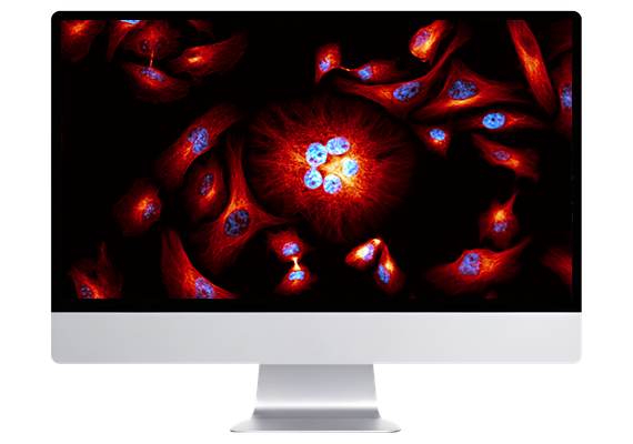
Imaris provides Core Facilities with a unified platform for visualising and analysing multidimensional imaging data from diverse microscopy systems. It helps standardise workflows across multiple users, improving consistency in analysis and reporting. The software supports both novice and experienced users through guided workflows and configurable tools. Its ability to process large datasets aligns with the demands of high-throughput imaging environments.
The power of Imaris for Core Facilities lies in our multiple detection models and the versatility they provide for all users and their research problems. Our 4 models (Spots, Surfaces, Cells and Filaments) can be harnessed to detect and analyse almost all biological samples, including cells, nuclei, nucleoli, bacteria, viruses, organs, neurons, dendritic spines, blood vessels and many more. Using them in combination alongside Imaris’ other tools opens the possibility to analyse dynamics of objects over time as well as their relationships with other objects in the image and to present these data in an elegant and informative fashion.
Deconvolution is often a first and important step in image analysis when users need to sharpen their images. Imaris overcomes common workflow problems where users need to open their files in various software packages, convert images and save multiple copies, by integrating deconvolution functionality into the Imaris menu. The main advantages of Imaris ClearView™ deconvolution are:
Stitching of individually acquired image tiles is an essential task for those who want to analyse big samples at high resolution. Imaris for Core Facilities includes Imaris Stitcher as most of Light-Sheet users and some Confocal Microscopy users aim to acquire these large images. Imaris Stitcher caters to these needs with an easy-to-use and performant interface. By including stitching functionality within the Imaris platform we’ve eliminated the time- and storage-consuming need for converting files from one format to another as users move between softwares.
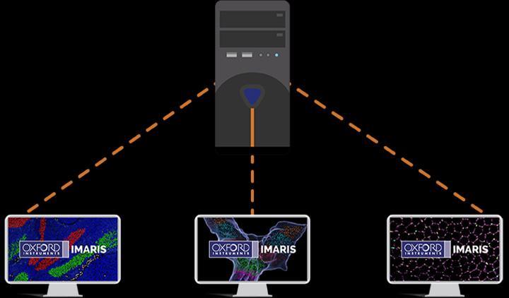
As a Core Facility Manager you may need greater flexibility than when a license is locked to one unique workstation so we have developed a Floating License Manager giving you the power to float the license to many different workstations within your lab, or department via your institution’s LAN. Expanded ranges beyond a department are available as well on request.
The Floating License Manager is also required when wishing to use Imaris on a workstation with app virtualization tools or where the workstation is running a server operating system. Purchasing one Imaris package license allows the use of Imaris on one workstation at a time. Additional licenses can be purchased if you are in a situation where multiple concurrent users are needed.
With Imaris 11, you can effortlessly create a customized image analysis protocol (Workflow) tailored to your specific research area and requirements. These Workflows can be saved and applied to other datasets with just one click, streamlining your analysis process. Discover how Workflows can accelerate your journey from microscopy data to meaningful insights, impactful publications, and engaging conference presentations.
The software supports a range of imaging workflows, from exploratory visualisation to advanced quantification and tracking. Its modular structure allows users to apply relevant tools depending on their experimental needs.

Users can adapt workflows to suit specific experimental needs. This flexibility allows facilities to support a wide range of applications without requiring separate tools. It helps maintain consistency while accommodating diverse research goals.

Imaris combines visualisation, segmentation, and quantification in a single environment. This reduces the need for multiple software tools and simplifies workflow management. It helps ensure consistency across users and experiments within core facilities.

Imaris supports analysis of 3D and time-lapse datasets across multiple channels. This enables comprehensive investigation of complex biological processes. It is critical for facilities working with advanced imaging techniques.
Cell Biology is areas of research in life sciences that focus on the fundamental processes of life. Cell biology encompasses a broad range of research areas and applications such as apoptosis, cell cycle & cell division, DNA damage, plant cell biology, vesicle trafficking, in vitro studies etc. As for model organisms, cell biology investigates them all, from the most simple prokaryotes (bacteria) to single-cell eukaryotes (yeast, fungus) and even multicellular organisms.
Basic cancer research combines several aspects of studying cancer cell phenotypes, gene expression and interactions with the microenvironment in vitro and in vivo to better understand carcinogenesis, malignancy and develop new potential therapies. Cancer research often requires applying advanced fluorescence microscopy to study cancer cell behaviour interaction with the environment and spatial distribution of the tumour models in a time lapse.
Developmental Biology are areas of research in life sciences that focus on the fundamental processes of life. Developmental biology studies the process by which multicellular organisms grow and develop. Research focuses on processes such: as metamorphosis, embryonic development, tissue growth, morphogenesis, stem cell differentiation, embryogenesis, plant development and regeneration. Research in these areas is done both on a microscopic and molecular level, and multiple technologies are needed to successfully accomplish this work.
Neuroscience is a multidisciplinary branch of science focused on the study of the nervous system and how the brain works. The field studies nervous system functions, brain function and the related structures such as the spinal cord. It combines anatomy, physiology, cytology, molecular biology, developmental biology and modelling in order to understand neurons and neuronal circuits. As neuroscientists often balance on the cutting edge of science, they require sophisticated methods such as fluorescence labelling, optogenetics, photostimulation and state of the art image analysis.
Automated classification of detected objects (cells, nuclei, vescicles) with a trainable Machine Learning Classifier (ML), based on selected statistic or the combination of 2 features. Classes are labelled and available for visual presentation, plotting and for downstream analysis (export of statistics).

© Oxford Instruments 2026
 Powered by Bioz