
Getting Started with Imaris
New to Imaris? Start with short tutorials covering navigation, visualization, image preparation, and basic object detection.
 Part of the Oxford Instruments Group

Imaris 11.1 introduces the new Imaris Training Hub, a built-in learning environment designed to help researchers get up to speed faster and become more independent with image analysis. From short tutorials and guided workflows to real datasets and application-focused examples, users can learn, practice, and explore without leaving Imaris. Whether you're completely new to image analysis or looking to tackle more advanced projects, the Training Hub helps you get to answers faster and with greater confidence.
Free Trial Request PricingAnyone running an imaging facility knows the challenge.
New users need help getting started. Researchers ask the same introductory questions. Staff spend valuable time explaining basic workflows instead of focusing on more complex scientific challenges.
The Imaris Training Hub was created to help change that.
By giving users access to practical learning resources directly inside Imaris, it becomes easier for them to learn independently, build confidence, and solve common analysis tasks on their own.
The result? Less time spent on repetitive training and more time devoted to impactful research.
View BrochureAccess tutorials, workflows, and example datasets that show not only which buttons to click, but also how to solve real image analysis problems.
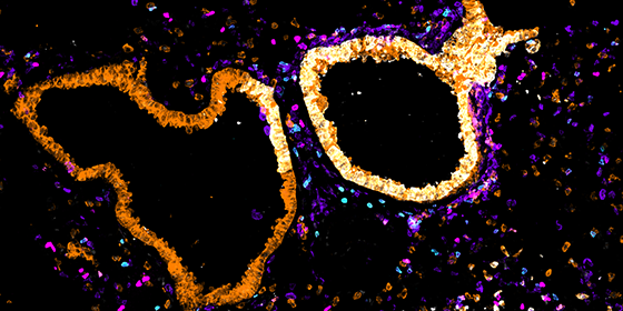
The Training Hub supports onboarding, self-learning, and workflow standardization, helping facilities scale training without scaling effort.
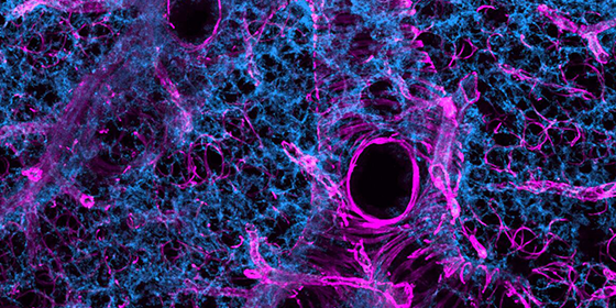
Shared workflows and practical examples make it easier to apply consistent analysis methods across projects, users, and experiments.
The Imaris Training Hub is organized around the way researchers actually learn and it is integrated directly into Imaris. Users can browse workflows, download datasets, watch tutorials, and access supporting materials without switching between websites, training portals, and documentation pages. Everything is available in the same environment where the work happens.

Getting Started with Imaris
New to Imaris? Start with short tutorials covering navigation, visualization, image preparation, and basic object detection.

Frequently Used Workflows
Step-by-step examples for common analysis tasks such as spot detection, surface creation, tracking, colocalization, and quantification.

Application-Focused Examples
See how Imaris can be applied to research questions in areas such as neuroscience, cell biology, live-cell imaging, and organoid research.

Advanced and Creative Workflows
Explore more specialized approaches for challenging datasets and unconventional analysis problems.
Imaris 11.1 also introduces several improvements designed to make everyday work easier.
New search and sorting tools in Imaris Arena make it easier to navigate datasets, workflows, videos, and documents.
Import and convert CZI datasets using the new built-in reader for a more streamlined workflow.
Technical and application support is now only a click away from within the software.
Access webinars, training events, and on-demand educational content through direct integration with Imaris Connect.
Imaris Viewer now allows users to view the workflows applied to a dataset, making it easier to understand how results were generated and shared across teams.
By bringing education directly into the software, we're helping users become productive faster while reducing the training burden on facilities. The goal is simple: make it easier for researchers to get reliable results and get more value from Imaris.
Anna Paszulewicz - Product Manager, Imaris
Cell Biology is areas of research in life sciences that focus on the fundamental processes of life. Cell biology encompasses a broad range of research areas and applications such as apoptosis, cell cycle & cell division, DNA damage, plant cell biology, vesicle trafficking, in vitro studies etc. As for model organisms, cell biology investigates them all, from the most simple prokaryotes (bacteria) to single-cell eukaryotes (yeast, fungus) and even multicellular organisms.
Basic cancer research combines several aspects of studying cancer cell phenotypes, gene expression and interactions with the microenvironment in vitro and in vivo to better understand carcinogenesis, malignancy and develop new potential therapies. Cancer research often requires applying advanced fluorescence microscopy to study cancer cell behaviour interaction with the environment and spatial distribution of the tumour models in a time lapse.
Developmental Biology are areas of research in life sciences that focus on the fundamental processes of life. Developmental biology studies the process by which multicellular organisms grow and develop. Research focuses on processes such: as metamorphosis, embryonic development, tissue growth, morphogenesis, stem cell differentiation, embryogenesis, plant development and regeneration. Research in these areas is done both on a microscopic and molecular level, and multiple technologies are needed to successfully accomplish this work.
Neuroscience is a multidisciplinary branch of science focused on the study of the nervous system and how the brain works. The field studies nervous system functions, brain function and the related structures such as the spinal cord. It combines anatomy, physiology, cytology, molecular biology, developmental biology and modelling in order to understand neurons and neuronal circuits. As neuroscientists often balance on the cutting edge of science, they require sophisticated methods such as fluorescence labelling, optogenetics, photostimulation and state of the art image analysis.

© Oxford Instruments 2026
 Powered by Bioz