Part of the Oxford Instruments Group
Expand

Imaris for Core Facilities

Imaris for Core Facilities is our recommended image analysis software package for microscopy facilities, larger labs and multi-user environments. Imaris is the market-leading visualization and analysis software for widefield, confocal (including spinning disk), light sheet, two-photon, electron microscopy, CLEM, OPT and many other modalities with over 4,000 publications in 2020 alone. Imaris for Core Facilities provides smart detection and visualisation of complex objects, tracing of neurons, blood vessels and other filamentous structures, 3D tracking (including cell division detection), interactive plotting and batch analysis.

Request Pricing/Free Trial Existing Customer - Satellite License

3D Microscopy Image Analysis

The power of Imaris for Core Facilities lies in our multiple detection models and the versatility they provide for all users and their research problems. Our 4 models (Spots, Surfaces, Cells and Filaments) can be harnessed to detect and analyse almost all biological samples, including cells, nuclei, nucleoli, bacteria, viruses, organs, neurons, dendritic spines, blood vessels and many more. Using them in combination alongside Imaris’ other tools opens the possibility to analyse dynamics of objects over time as well as their relationships with other objects in the image and to present these data in an elegant and informative fashion.

Spots

Surfaces

Cells

Filaments

Quantitative Image Analysis for 3D Microscopy

After detecting and segmenting the image data as objects using one of the 4 models, Imaris calculates a wide range of statistics. All values can be used for, filtering, labelling, color coding, plotted inside Imaris (using Vantage plots) or exported in a preformatted .csv or .xls file. Machine Learning Object Classification tool uses many of these values in addition to many more machine learning specific ones. Although the parameters presented below are the most common statistic types needed by biologists, Imaris reports many more.

Motion Analysis
  • Speed
  • Acceleration
  • Cell division tracking
  • Trajectory
  • Event synchronization
Quantification
  • Count of objects
  • Area
  • Volume
  • Intensity
  • Position
Interactions
  • Distances between structures
  • Volume overlap, contacts
  • 3D intensity profiling
  • Spatial distribution
  • Co-localization
Batch and Plots
  • Image processing
  • Measurements
  • Interactions
  • Group comparisons
  • Statistical tests
Filaments/Neurons
  • Length, Straightness
  • Mean Diameter
  • Branching Angle
  • Spine Density
  • Spine Shape
Cells
  • Volume
  • Vesicles per Cell
  • Distance to Membrane
  • Vesicles per Nucleus
  • Morphology

Image Analysis for Everyone

All users of your facility will experience the simplicity of the Imaris software while using our wizard-driven smart workflows and software hints. To improve the learning curve and facilitate the usage of our software the Imaris team has prepared full online introductory training: Image visualization and analysis in Imaris – Imaris Homeschool and numerous short video tutorials.

As an additional service the Imaris Team can provide customized trainings for all lab members or facility users. Trainings can be delivered on-line (via Amazon Web Services) or on-site at user’s facility.

For personalized workflows (help on specific applications) and assistance you can always contact image analysis and life sciences specialists from the Imaris’ Technical and Application Support Team (included in Imaris Maintenance plan). We encourage all users to let our team of experts assist you in getting the most out of their images.

To help your users share their data with collaborators or visualise their data at home or conference we offer Imaris Viewer – free software which enables opening both native microscopy files and datasets already analysed in Imaris.

Flexible Licensing Options

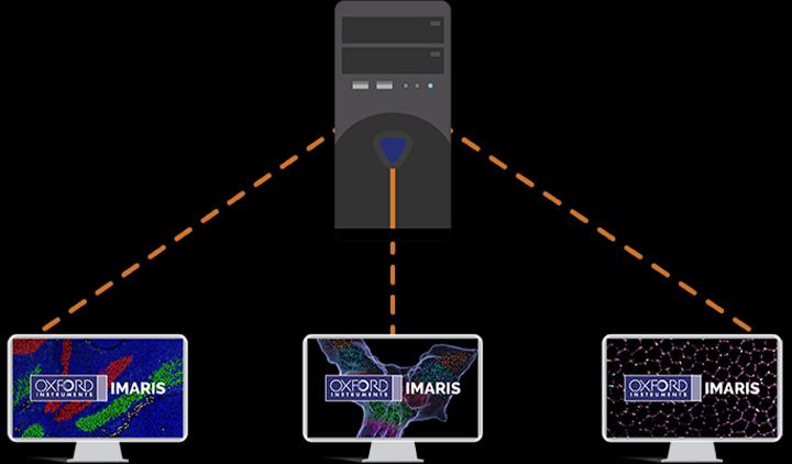
As a Core Facility Manager you may need greater flexibility than when a license is locked to one unique workstation so we have developed a Floating License Manager giving you the power to float the license to many different workstations within your lab, or department via your institution’s LAN. Expanded ranges beyond a department are available as well on request.

The Floating License Manager is also required when wishing to use Imaris on a workstation with app virtualization tools or where the workstation is running a server operating system. Purchasing one Imaris package license allows the use of Imaris on one workstation at a time. Additional licenses can be purchased if you are in a situation where multiple concurrent users are needed.

ClearView™ GPU-accelerated Deconvolution

Deconvolution is often a first and important step in image analysis when users need to sharpen their images. Imaris overcomes common workflow problems where users need to open their files in various software packages, convert images and save multiple copies, by integrating deconvolution functionality into the Imaris menu. The main advantages of Imaris ClearView™ deconvolution are:

Stitching of big image tiles for Light-Sheet and Confocal Microscopy

Stitching of individually acquired image tiles is an essential task for those who want to analyse big samples at high resolution. Imaris for Core Facilities includes Imaris Stitcher as most of Light-Sheet users and some Confocal Microscopy users aim to acquire these large images. Imaris Stitcher caters to these needs with an easy-to-use and performant interface. By including stitching functionality within the Imaris platform we’ve eliminated the time- and storage-consuming need for converting files from one format to another as users move between softwares.

Users will benefit from:

Additional Resources

The Imaris Learning Center hosts a wide range of tutorial videos, how-to articles and webinars to guide you through the many features of Imaris. We have provided some links below which will get you started on some of our most recent developments.