Part of the Oxford Instruments Group
Expand

Cancer Research

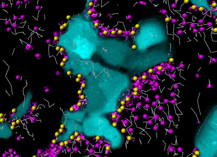
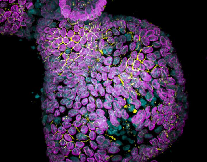
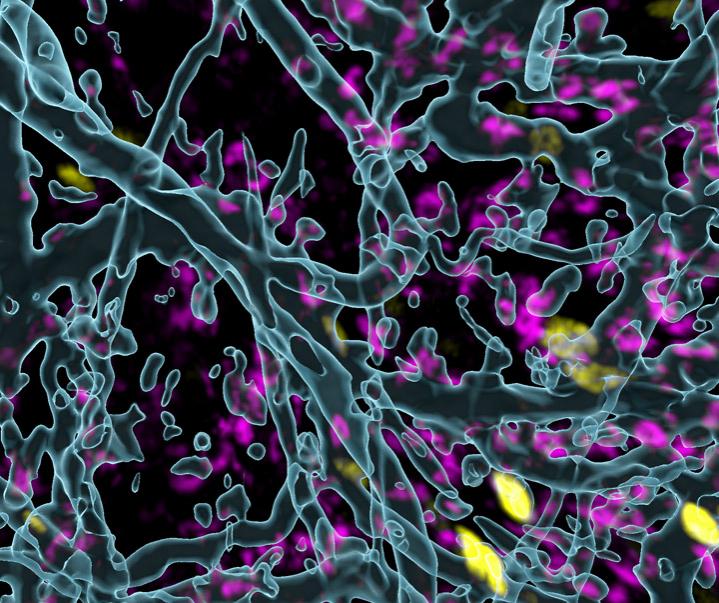
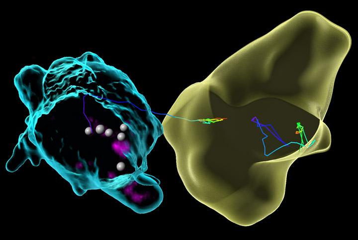
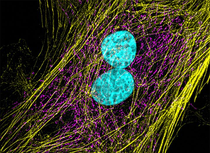
Basic cancer research combines several aspects of studying cancer cell phenotypes, gene expression and interactions with the microenvironment in vitro and in vivo to better understand carcinogenesis, malignancy and develop new potential therapies. Cancer research often requires applying advanced fluorescence microscopy to study cancer cell behaviour interaction with the environment and spatial distribution of the tumour models in a time lapse. Learn about Andor solutions for high-speed and sensitive image acquisition and Imaris for 3D quantitative image analysis, working together for faster discoveries in cancer research.

Request Pricing

Applications and Techniques

Immunotherapy

Cancer immunotherapy employs human own immune systems to attack cancer cells and has become a promising modality of cancer treatment based upon the clinical successes of immune checkpoint blockade and adoptive T cell transfer. The success of immunotherapy of cancer depends on several cellular events in the tumors that can be visualized by live microscopy strategies in experimental models. Intracellular events can be captured by fluorescence microscopy on living cocultures of T cells and tumor cells. 

Dragonfly spinning disk confocal equipped with back-illuminated Sona sCMOS allows for fast acquisition and low phototoxicity is an excellent solution for imaging the interactions between immune and cancer cells because it allows for long exposure studies. Imaris for Cancer Research package allows researchers to analyse the dynamic events occurring in the experimental setup, such as tracking, volume changes or behaviour after cell contact.

Organoids

The 3D cell culture technologies, such as organoids can act as human cancer models in near physiological conditions. Such preclinical organoid models are the bridge between basic cancer research and testing therapies for patients with cancer. Using organoids researchers can also model infection-cancer progression and mutation-carcinogenesis processes. In addition, organoids can open door to personalized cancer treatment regimens as they can be grown with high efficiency from patient-derived healthy and tumour tissues.

Confocal imaging devices are necessary for in-depth evaluation of organoid morphology and behaviour. Dragonfly spinning disk confocal is a system that provides flexibility in imaging magnification, illumination, and mode while also improving data acquisition speed. Imaris for Cancer Research and Imaris for Cell Biologists image analysis packages offer wide range of tools for 3D image visualization, segmentation, and detailed quantitative measurements to understand the organoid models.

Tumor Microenvironment

Studying tumor requires visualization of its microenvironment including blood vessels, extracellular matrix and immune cells. Tumor constantly interacts with its surrounding and imaging it puts light on the complex interaction between various elements, which may lead to better understanding of cancer progression and response to therapeutics. To study those interactions, researchers need to acquire 3D volumes, often time-lapse movies that show tumor cells at the single cell level as well as other features from the tumor microenvironment such as the blood vessels. 

The perfect solution for intravital in-vivo fluorescence imaging of such samples is multiphoton microscopy which allows for deep tissue penetration, equipped with fast and sensitive back-illuminated Sona sCMOS or iXon EMCCD. Imaris for Cancer Research enables 3D multicolor visualization of complex datasets, segmentation and tracking of objects of interest to study the subtle dynamic changes of the system, hence is the perfect image visualization and analysis solution tool for studying tumor in its natural microenvironment. 

Spatial Transcriptomics

Spatial transcriptomics in cell biology is the detection of numerous (Xn) RNAs (or other biological molecules) in a tissue /cell, either in its 2D or 3D context. It can appear under many names in the literature: spatially resolved transcriptomics, spatial transcriptomics, multiplexing, and in-situ multiplex imaging. The key advantage of spatial transcriptomics is its ability to help understanding where genes are expressed and their surrounding environment, or microenvironment.  In cancer research this powerful technique can help predict the evolution of the disease due to the identification of key expression genes. 

Dragonfly is an excellent solution for in-situ multiplexing hybridisation. Dragonfly spinning disc confocal with large field of view allows for rapid acquisition of tissue volumes. Additionally, the dual microlens disk system combined with Andor´s high dynamic range and high QE cameras such as back-illuminated Sona sCMOS or iXon EMCCD series allows the capture of all the signals in the sample: from the dimmest to the brightest.

Contact Application Specialists

Cancer Cell Motility 

Cancer cells are known to be more motile than the normal cell and this ability contributes to tumour metastasis events. Metastasis is one of the greatest challenge in cancer therapies leading to faster and uncontrolled disease progression. Studying cell motility and identification of genes responsible for the enhanced motility of cancer cells  can lead to better understanding of cancer invasion and metastasis and eventually development of more efficient therapeutic strategies for patients with cancer.

Dragonfly spinning disk confocal with fast and sensitive back-illuminated Sona sCMOS is a perfect solution for live cell imaging and migration studies. Imaris for Cancer Research enables tracking of cells including cell divisions over long periods of time and analysing motion parameters, such as speed, displacement and many more.

Cytoskeleton Studies

Studies involving cytoskeleton under normal and pathological conditions are crucial to understand cancer cell behaviour. It has been demonstrated in several studies that normal cell behaviours involving cytoskeleton are altered in cancer cells. Due to their vital role in cell movement, cytoskeleton components: actin filaments and microtubules have become a target of several anticancer therapies. Examining how cancer cells use cytoskeletal proteins to move through the body may lead to targeted therapies that reverse these protein signals.

Due to its size, cytoskeleton studies require precise confocal microscope equipped with super-resolution capabilities such as SRRF-Stream+ compatible iXon Life/iXon Ultra EMCCD or Sona back-illuminated sCMOS. Imaris for Cell Biologists is a perfect image analysis package to study cytoskeleton and its interaction with other cell components.

Solutions for Cancer Research

Learning Center Resources

Publications