Part of the Oxford Instruments Group
Expand

Cell Biology

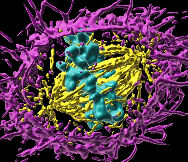
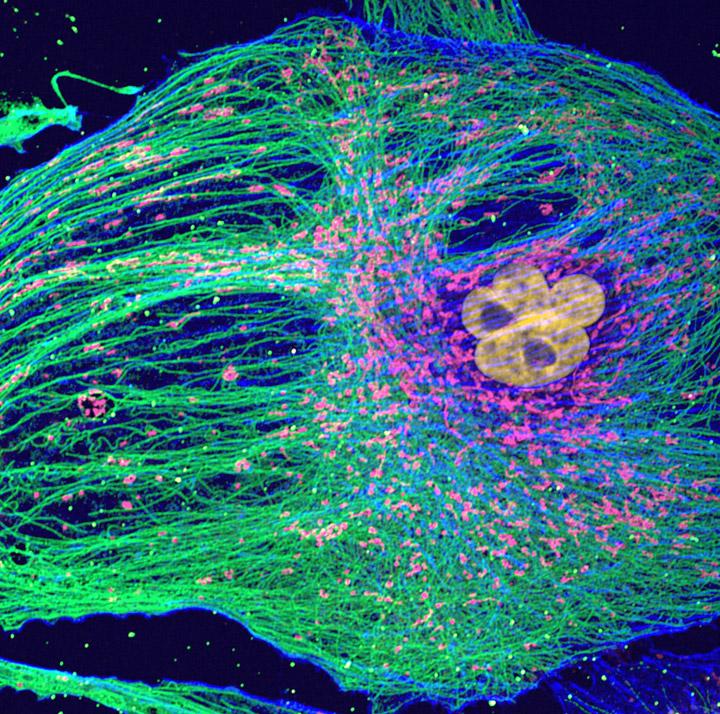
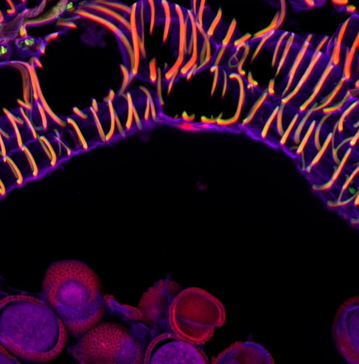
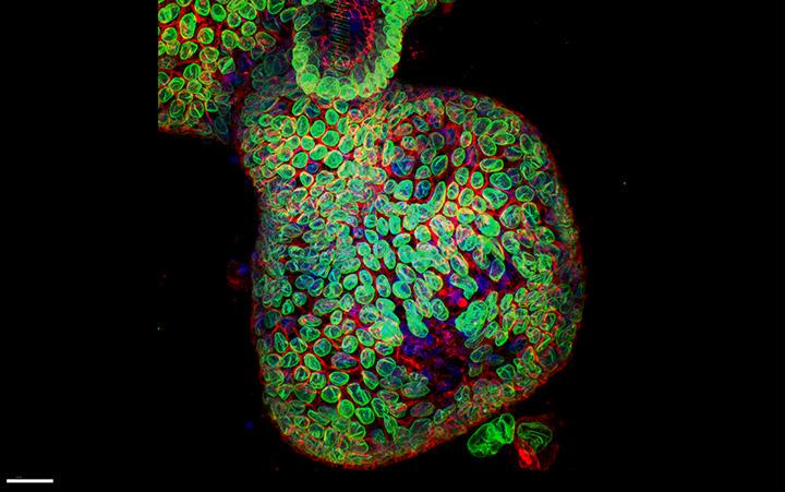
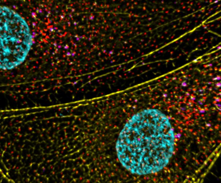
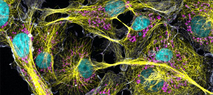
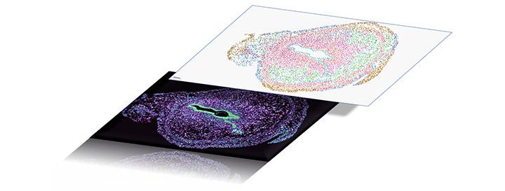
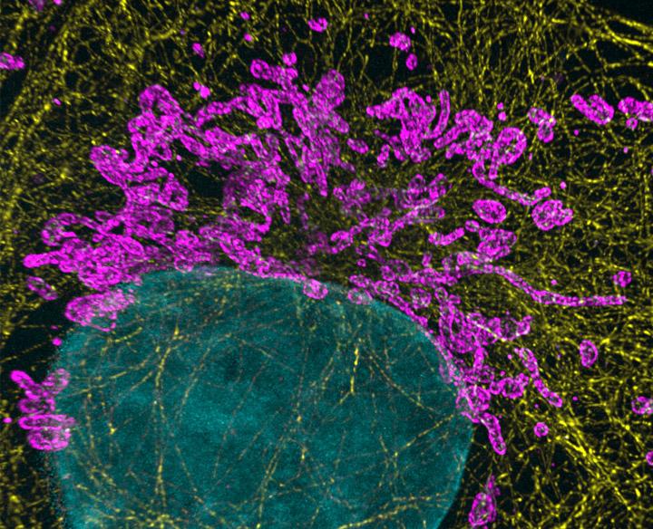
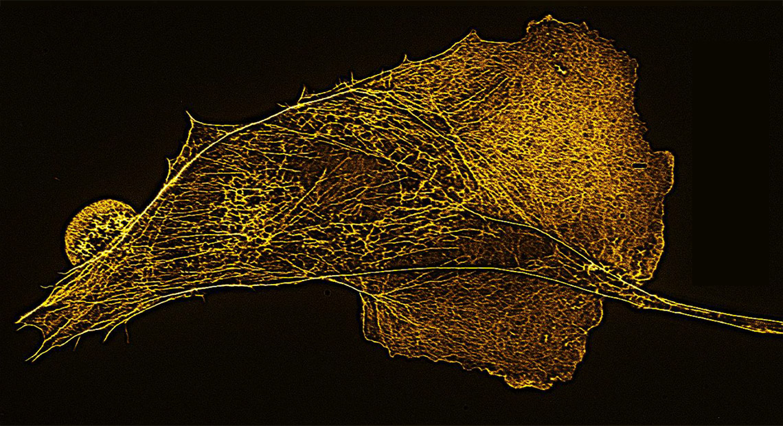
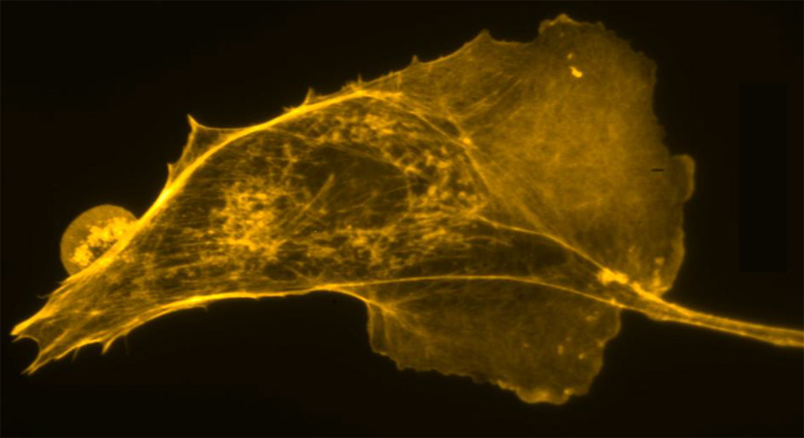
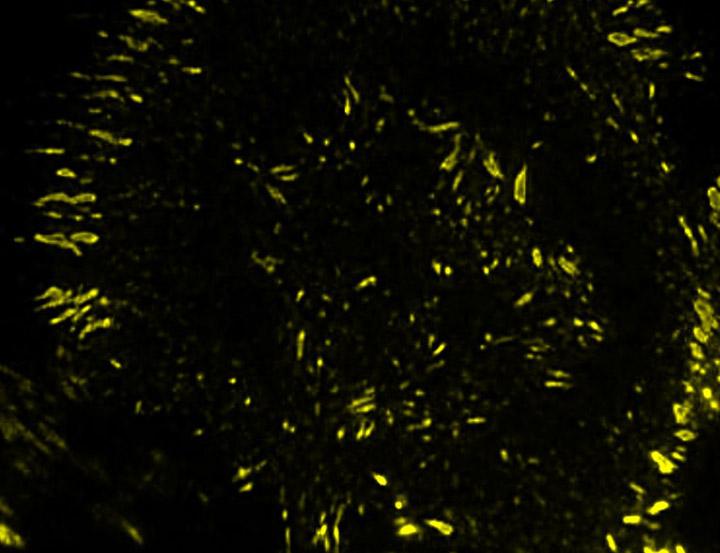
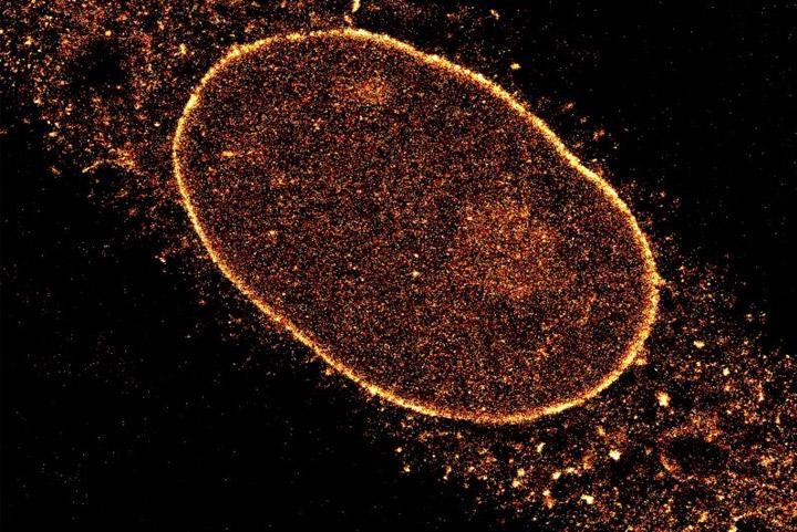
Cell Biology is areas of research in life sciences that focus on the fundamental processes of life. Cell biology encompasses a broad range of research areas and applications such as apoptosis, cell cycle & cell division, DNA damage, plant cell biology, vesicle trafficking, in vitro studies etc. As for model organisms, cell biology investigates them all, from the most simple prokaryotes (bacteria) to single-cell eukaryotes (yeast, fungus) and even multicellular organisms. Andor provides the technological solutions to tackle cell and developmental biologists research challenges.

Request Pricing

Applications

Cell Migration

Cell migration includes the study of cytoskeletal dynamics and the membrane morphology of moving cells. Cytoskeletal dynamics and membrane morphology of moving cells need to be imaged with high resolution and sensitivity with minimal phototoxicity and photobleaching. 

For extremely light-sensitive thin samples such as single cells or tissues slices both the Andor Benchtop Confocal and Dragonfly in widefield imaging mode are ideal choices. Using Andor Dragonfly, the researchers can further image extremely high-speed dynamic events such as cilia imagining (>50 fps). The TIRF mode can be used for high-resolution membrane-substrate interactions (adhesion studies). Both confocal systems allow imaging of thicker low signal-to-noise samples. 

In addition, analysis of cell movement events benefits from EMCCD sensitivity to allow very low light imaging or sCMOS technology to capture high-speed dynamic events such as cilia beating. 

Andor Mosaic can be used as an optogenetic to study cytoskeleton dynamics in motile cells. Furthermore, Imaris for tracking is the ideal solution for automatically analysing moving objects over time.

Contact our Application Specialists

Mitosis & Meiosis

Mitosis and Meiosis are the processes that define cell division. Mitosis is the division of somatic cells. Meiosis is the division of germ cells to originate the gametes. Both mitosis and Meiosis are dynamics event and be analysed live and fixed.

Operating Andor Benchtop Confocal and Andor Dragonfly  widefield mode allows image mitosis in monolayer cells under very low light conditions. The confocal is the ideal choice for thicker samples such as c. elegans. Imaging cell division along a large tissue area will benefit from sCMOs large field of view. For live super-resolution applications such as kinetochore-microtubule interaction, Dragonfly equipped with EMCCD and SRRF-Stream licence is the ideal choice.

Andor's Mosaic will allow users to specifically activate a subset of chromosomes, kinetochores and microtubules while mitosis is proceeding. For laser ablation of mitotic structures as the centrioles and kinetochores, MicroPoint is your choice.

With Imaris for Cell Biologists, a multitude of analysis tools are available to create 3D renderings of mitosis as well as to plot the analysis of events such as chromosome congression or metaphase to anaphase transition timing.

Organelles Biology

Organelles are subcellular structures delimited by a membrane, which are specialized in specific tasks in the eukaryotic cell. Organelles include the nucleus, mitochondria, endoplasmic reticulum, chloroplasts, etc.

Andor Dragonfly and highly sensitive camera Ixon EMCCD are ideal for studying organelle bidirectional movements such as mitochondria and lysosome movements. For vesicle trafficking and fusion events, Andor Dragonfly coupled with the small pixel size Sona and SRRF-stream licence will deliver the resolution required for such experiments. Fast imaging of calcium waves can be captured with Andor Benchtop Confocal. Ultra-fast imaging of Endoplasmic Reticulum-calcium signalling can be captured with Dragonfly coupled to a Sona/Zyla sCMOs camera.

Further, the detailed structural analysis of 3D organelles such as mitochondria using 3D-dSTORM the solution is imaging with Andor Dragonfly and a Sona 6,5 micron Pixel camera.

Photoactivation of inner Golgi vesicles with Mosaic allows following vesicle trafficking of targeted events. As for nuclear DNA damage studies, MicroPoint is the ideal tool.

For data analysis, Imaris for Cell Biologists allows the quantification of organelle bidirectional movement, intra-organelle distance, also delivering spectacular 3D renderings and movies of the data.

Plant Cell Biology

Plant cell biology focus on a broad area of research in which the model organism are plants or plant cells. The area encompasses various subjects such as cellular structures and function, molecular and cellular mechanisms, inter-organelle communication and intracellular signalling, etc.

Andor offers a broad range of solutions for plant cell biologists. The Andor benchtop confocal is the ideal choice for more typical imaging applications such as live-cell imaging and fixed tissue sample analysis. When imaging high background plant samples, researchers can take advantage of the Andor Dragonfly extended spectral range with excitation wavelengths up to 750 nm. TIRF imaging is ideal for minimising the high background (high autofluorescence) associated with plant tissues; it can be powerful when analysing membrane fusion events in plant cells. The detailed structural organisation of cortical microtubules in plant roots can be visualised with dSTORM. For live-cell super-resolution of endosomes and plasma membrane dynamics, Andor Dragonfly equipped with Sona 6,5 with SRRF-stream licence will be the ideal solution. Both the iKon-M CCD camera and the Ixon EMCCD deliver excellent results for plant Bioluminescence applications such as Circadian Clock regulated transcription.

Imaging analysis with Imaris Essentials allows the researchers to have an interactive visualisation of 3D snapshot and time-lapse images as well as generate quantitative information from the microscopy data.

Stem Cell 

Stem cell biology has a multitude of research areas, from pluripotent stem cells to organoids, from neurosciences stem cells to cancer stem cells, etc. 

Due to their high background rejection Imaging deep into steam cell generated organoids can be accomplished with Andor Benchtop Confocal and Dragonfly. Further, the large field of view delivered by Andor confocal microscopes results in a massive increase in productivity. 

In addition, dragonfly, in combination with sCMOS cameras, delivers allows to image highly dynamic processes as, for example, intracellular transport in organoids. 

For cancer embryonic stem cells, as for organoid stem cells, a significant challenge is the penetration deepness. Using Dragonfly NIR lasers and NIR sensitive cameras (EMCCD or sCMOS) will increase the optical penetration considerably with minimal background. 

Andor Mosaic will be the ideal tool to track engineered stem cells expressing light-activated opsins, allowing to track the differentiation of the neuronal stem cells. 

As for image analysis and data presentation, Imaris for Cancer Research will allow following the lineage of cancer steam cells; Imaris for Neuroscientists allow to track filaments in neuronal stem cells. Finally, Imaris for Cell Biologists allows to segment the cells and different compartments.

Contact our Application Specialists

Vesicle Trafficking

Vesicle trafficking is a significant area of research in cell Biology; it extends from cell membrane to organelles and ensures cell communication with the external environment.

Andor offers a broad range of solutions to tackle vesicle trafficking challenges. For cell surface events of endo and exocytosis, using Andor Dragonfly in TIRF mode coupled to a Sona 6,5 sCMOS camera will deliver maximum resolution. Dual camera simultaneous imaging option allows simultaneous observation of the different players in the endocytic event, as the constituent of clathrin-coated vesicles (dynamin, AP-2, actin fibres, …). To follow the endocytic from membrane fusion till late lysosome formation process inside the cell with high temporal and spatial resolution, researchers can take advantage of Dragonfly speed coupled with SRRF-Stream for live-cell super-resolution. The extreme sensitivity of our iXon EMCCD cameras will allow very low light imaging, dramatically minimising phototoxicity.

Imaris for Cell Biologists allows automatic detection and quantification of vesicles in 3D over time.

Techniques

Confocal Microscopy

Confocal spinning disk microscopy is ideal for multiple cell biology applications; it delivers gentle imaging for live cell experiments such as mitosis. It allows fast 3D scanning for samples such as stem cell-derived organoids.

With a spinning disk confocal microscope, images of fixed or live cells can be acquired, and the researcher can have an insight into the cell organelles 3D spatial organization.

Multiplex Imaging

Spatial transcriptomics (or Multiplexing) in cell biology is the unveiling of several (Xn) RNAs in its 2D or 3D biological context. The advantage of spatial transcriptomics is its ability to understanding where genes are expressed and their surrounding environment in multiple gene products.

Mechanistically, fluorescent probes label the hybridised RNA molecules, the image data is acquired (typically a volumetric montage is scanned), and the probes are washed away. After each image dataset is acquired, a “strip and wash” step is followed by another hybridisation round. This procedure is repeated N times and results in large volumes of encoded image data. A highly sensitive spinning disk microscope and camera with large Filed of view, that deliver uniform illumination across the whole imaging field is the ideal choice for multiplex imaging. 

Expansion Microscopy

Expansion Microscopy (ExM) is an imaging protocol that delivers super-resolution information about the sample being analysed. In expansion microscopy, instead of optically breaking the diffraction limit of light (<200 nm), the super-resolution is achieved by isotropically expanding the sample. This technique allows conventional light microscopes to see sub-diffraction limited or densely packed details that previously could not be distinguished in the light microscope.

Expanded samples are quite large, and to increase productivity microscope with a large field of view is ideal for visualising expanded samples. ExM images also benefit from Borealis uniform illumination to seamlessly merge all the captured tiles as well as of highly sensitive EMCCD detectors. Mitochondria, centrioles peroxisomes, and nucleus are organelles whose ultra-structure can be image using Expansion microscopy.

SRRF-Stream

SRRF-stream is an alternative approach to super-resolution. SRRF-stream can be combined with any imaging modality (e.g. widefield, TIRF or confocal), and the final resolution will depend on the proprieties of the acquired data sets. With SRRF-stream, the researchers can achieve resolutions in XY up to 50 nm. SRRF is a live cell imaging compatible super-resolution technique that does not require specific sample preparation, that can deliver super-resolution images as fast as 10 frames per second (depending on the acquisition devices) on the fly. 

To visualise mitochondria or combine with TIRF to increase the resolution in imagining the membrane fusion events, SRRF will deliver resolutions beyond the diffraction limit. Importantly, SRRF-Stream is compatible with confocal imaging and can provide super-resolution images deep inside cells and tissues when combined with DragonFly spinning disk confocal imaging.

After
Before
F-actin of BPAE cells labelled with Alexa Fluor 488 Phalloidin imaged on a Nikon Ti2 microscope at 60x and Sona 4.2B-11.

To visualise mitochondria or combine with TIRF to increase the resolution in imagining the membrane fusion events, SRRF will deliver resolutions beyond the diffraction limit. Importantly, SRRF-Stream is compatible with confocal imaging and can provide super-resolution images deep inside cells and tissues when combined with DragonFly spinning disk confocal imaging.

TIRF Microscopy

A TIRF microscope (total internal reflection fluorescence) allows the user to acquire very detailed images of an object at the cell surface. TIRF relies on differences between the refractive index of the medium where the sample is inserted and the refractive index of the glass slide.

One consequence of this type of illumination is that images can only be acquired at a very thin boundary between the Interface of the two different media. TIRF will allow imaging up to a maximum of 100-200 nanometres inside the sample. Thus, TIRF is the ideal solution for analysing live cell events at the cell membrane boundary, such has, membrane dynamics, vesicle trafficking, endocytosis, exocytosis and any other events at the cell surface. Furthermore, the high resolution provided by a TIRF system makes it also a valuable technique for single-molecule imaging.

Single Molecule Localization Microscopy

Super-resolution microscopy has allowed a deeper understanding of the cell. SMLM (Single Molecule Localization Microscopy) delivers resolutions up to 20 nm laterally. The use of an astigmatic lens allows a calibrated distortion on the PSF that will deliver axial (Z) information, delivering 3D single-molecule localization.

dSTORM relies on acquiring images of the Fluorophores that switch between the ON and OFF states. On the other hand, DNA-PAINT does not require photobleaching; it relies on the transient immobilization of fluorophores to emit the signal. The immobilization is achieved by the hybridization of DNA strands. SMLM microscopy using DNA-PAINT benefits from acquiring images in the TIRF modality to reduce the background

Solutions for Cell Biology

Learning Center Resources

Customer Publications