Part of the Oxford Instruments Group
Expand

Developmental Biology - Microscopy Imaging Solutions

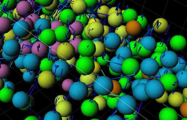
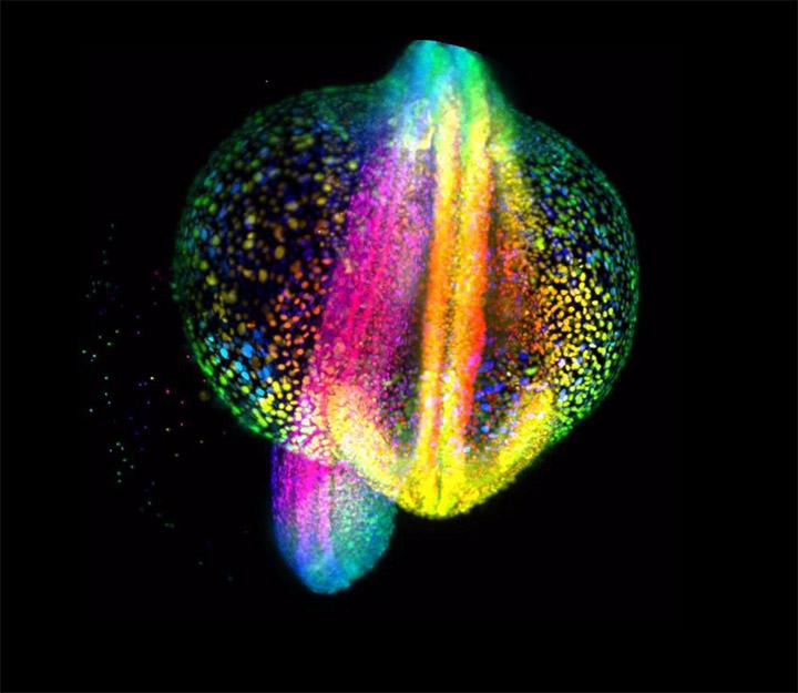
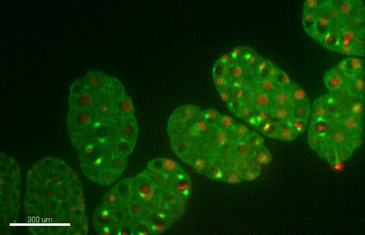
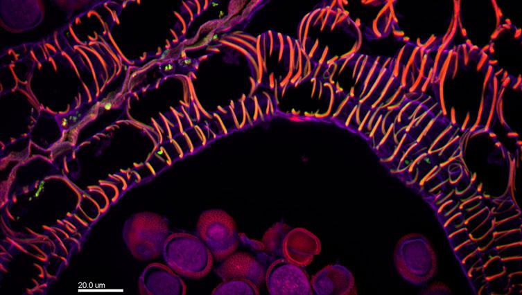
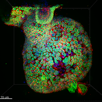
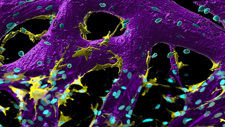
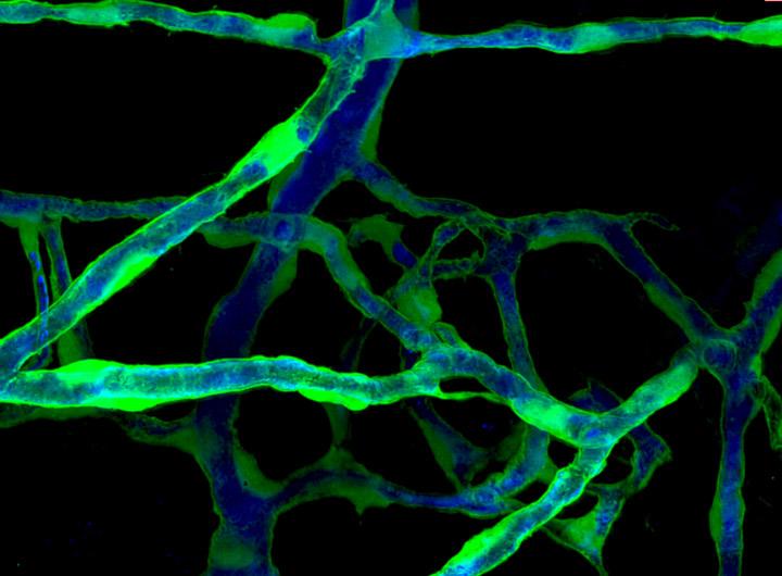
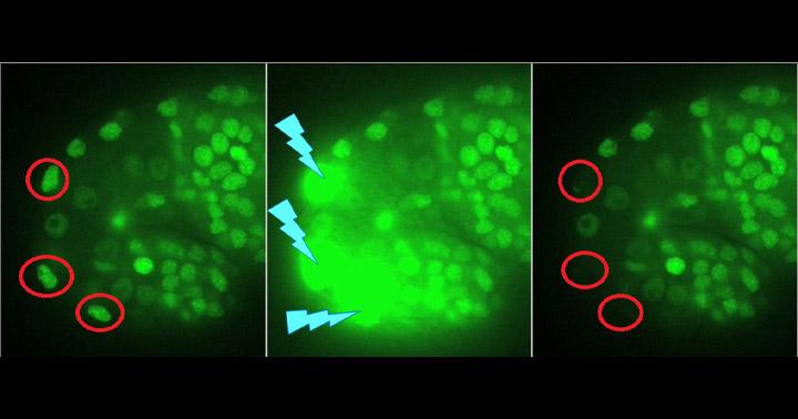
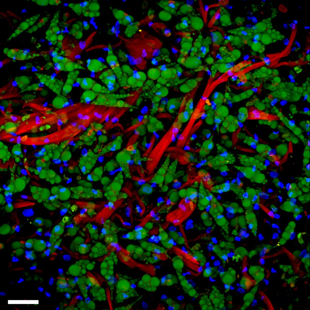
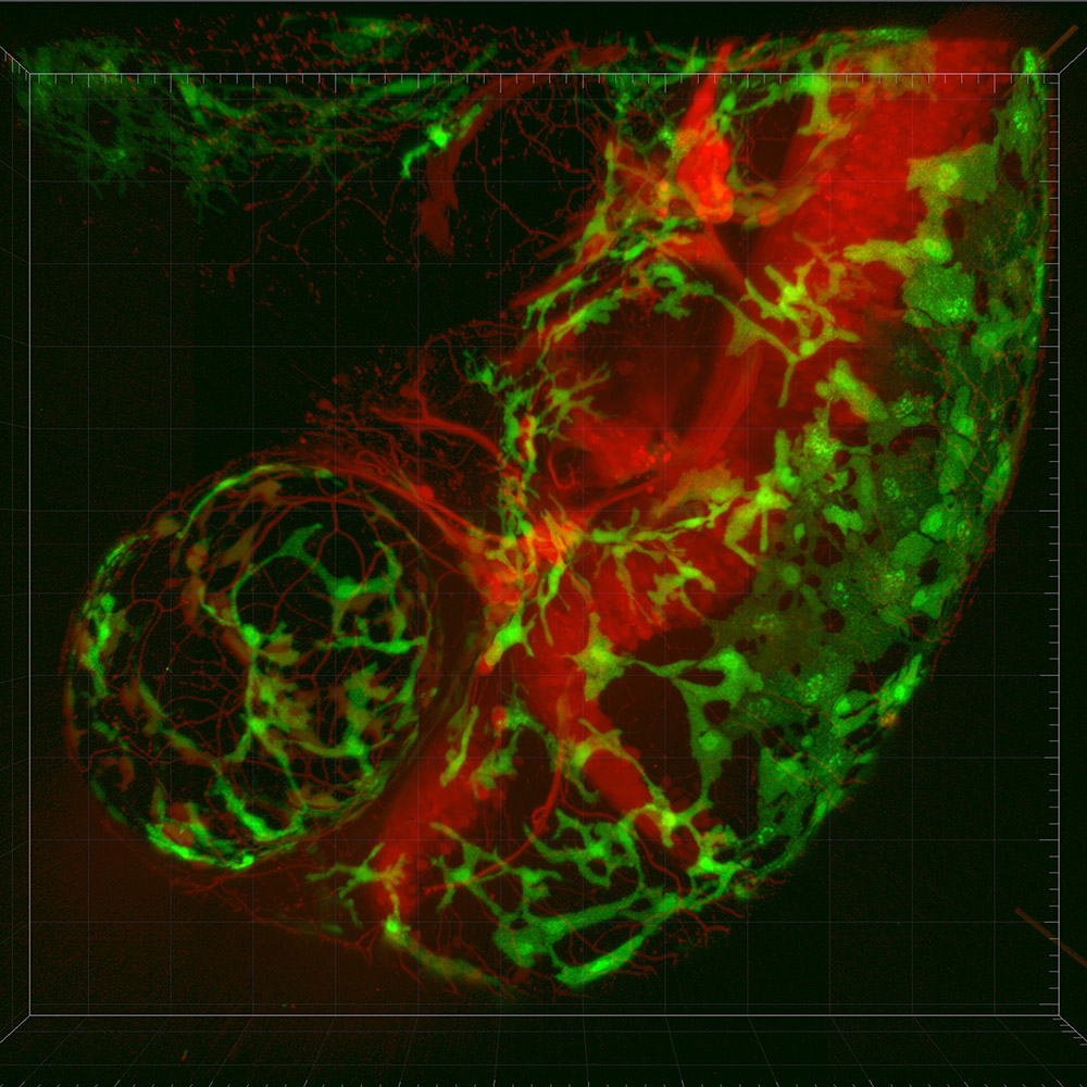
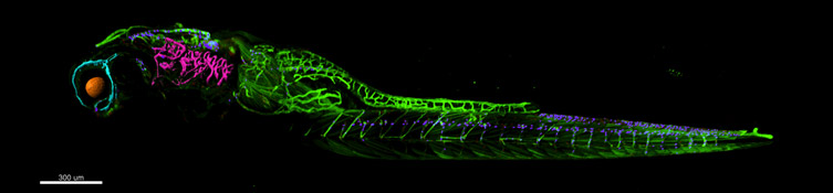
Developmental Biology are areas of research in life sciences that focus on the fundamental processes of life. Developmental biology studies the process by which multicellular organisms grow and develop. Research focuses on processes such: as metamorphosis, embryonic development, tissue growth, morphogenesis, stem cell differentiation, embryogenesis, plant development and regeneration. Research in these areas is done both on a microscopic and molecular level, and multiple technologies are needed to successfully accomplish this work. Imaris provides the technological solutions to tackle Cell and Developmental Biologists research challenges.  

Request Pricing

Applications

Cell Migration

In developmental biology, cell migration is a crucial aspect of research. Cells can migrate independently or collectively. Collective cell migration ensures the maintenance of cell-cell contacts, organism group polarisation and is orchestrated in a coordinated way. 

Andor offers the solution to analyse the dynamics of cell movement in real-time. The high background rejection and sensitivity of Dragonfly, as well as the large Field of View offered by Andor dragonfly and sCMOS camera (such as Sona 11), ensure that collective cell migration can be imaged throughout days without phototoxicity or photobleaching. Using Imaris for tracking, the researcher can automatically analyse moving objects, statistics and plot the results. Importantly this data will be accompanied by stunning 3D rendering images.

Contact our Application Specialists

Morphogenesis

Morphogenesis researchers study the molecular and cellular mechanisms that drive the development of an entire organism. It can also be studied in a mature organism focusing on tissue homeostasis, tissue regeneration and cancer cell morphogenesis.

Imaging live morphogenesis requires a highly sensitive microscope that can deliver images with extremely low light. Dragonfly combined with the high sensitivity on an EMCCD camera is the ideal choice to study morphogenic dynamics.

To address how gene expression affects morphogenesis, the Andor cryostat, the MicrostatN, or the Microstat He-R are ideal solutions to cut the sample with high precision. To ensure high productivity, image acquisition should be done, taking advantage of the high speed of Andor Spinning Disk confocals (BC43 or  Dragonfly). A large sensitive and Field of View sCMOS camera is also an important choice.

The image analysis will depend on the research topic:  Imaris for Tracking allows the tracking to track cell lineage, and Imaris for Cell Biologists can address colocalization of gene expression.

Contact our Application Specialists

Embryogenesis

Embryogenesis is the development of an organism from egg to completion of the embryonic stage. 

Both Andor Benchtop and Dragonfly confocal microscopes are excellent tools for studying embryogenesis due to the large Field of View. Researchers can capture entire developing embryos from single-cell state to organogenesis. 

Dragonfly near-infra-red (NIR) lasers are an outstanding option for imaging live imaging the developing embryo. Address the mechanisms of asymmetric cell division with minimal light intensity using Dragonfly coupled to an EMCCD camera

With Andor Benchtop confocal, image early embryonic development of Zebrafish (or other large model organisms). Visualise the whole organism with DPC and localise the fluorescence by combining DPC with confocal imaging. 

Use optogenetics tools to address the fate of specific cell populations (Andor Mosaic). Local cell movements can be induced with stronger lasers pulses (Micropoint). Activate or sequester specific proteins and analyse their influence on meso, ecto and endoderm formation. 

For tracking cell lineages and cell movement through embryogenesis, analysing different cell subpopulations or even plotting the distribution of embryonic gene expression markers, Imaris for Tracking and Imaris for Cell Biologists provide the researchers with the necessary tools for image analysis quantification and 3D rendering

Contact our Application Specialists

Plant Research

The number of questions posed to developmental plant biologists are enormous.

For Gene expression analysis aiming at generating a database of cell types and genome specificity, the sample needs to be sliced. MicrostatN, or the Microstat He-R, are ideal solutions to cut plant tissues.

The large Field of view of Andor Dragonfly combined with patented Uniform Borealis Illumination is ideal for imaging multiple colour plant samples. Back Illuminated sCMOS cameras can deliver the speed and the Field of view required to speed up the capture of the vast amount of experimental data. NIR lasers are ideal for imaging deep into sample tissues cutting through the troubles of scattering and autofluorescence.

Andor dragonfly and the Ixon EMCCD camera with SRRF-stream tackles the challenges of speed and diffraction-limited resolution in plant vesicle trafficking.

Due to their low noise on very long exposures, Ikon cameras are ideal for Bioluminescence studies, whereas IXon EMCCDs are ideal for single-molecule bioluminescence imaging due to high sensitivity.

Imaris for Cell Biologists allows the segmentation of different plant tissues and the segmentation of intracellular plant structures such as cell wall, cell membrane, nucleus, chloroplast, etc. In addition, the 3D rendering images can be accompanied by graphics that plot intercellular /intra-organelle distance in XYZ and other statistical measurements.

Contact our Application Specialists

Organoids

Organoids are 3D structures that can replicate the complexity of an organ, and they are one of the most promising and most rapidly evolving tools in scientific research.

Due to its large field of view, uniform Borealis illumination, and acquisition speed, both Andor Confocal systems Andor Benchtop and Andor Dragonfly excel in imaging 3D organoids.  The Andor Benchtop is the best option for samples with four colours and up to 500 mm thick. For deeper imaging the researchers should choose Andor Dragonfly.

The large field of view and high dynamic range delivered by sona 4.2-11 is ideal for capturing all the emitted light  (high and low intensity) from the organoids.

 Low light live imaging of steam cell differentiation into organoid formation will require a highly sensitive camera such as the iXon EMCCD.

Optogenetics/photostimulation experiments can address the cell fate at the development from stem cell to an organoid. Mosaic is the ideal tool to locally activate one or more cells in multiple regions of the developing organoid.

As for protein recovery dynamics at specific cellular structures or defined regions of the organoids such as cell membrane, laser irradiation with Micropoint to FRAP-bleach the desired area will be the ideal solution.

Imaris for Cell biologists delivers all the tools required for organoid biologists, from tracking cell movements to identifying different cell populations and linage. All are available combined with robust statistics and stunning 3D rendering movies and images.

Contact our Application Specialists

Vascular Research

Vascular research addresses how the blood vessels are formed and how the blood flows in the arteries and veins. It has applications from addressing vascularization on organ/organism development to cancer.

The Andor cryostats, the MicrostatN, or the Microstat He-R, are ideal solutions to cut the sample with high precision and address the blood vessel wall formation in fixed samples.

Andor high-speed multipoint confocal systems are coupled Borealis uniform illumination, which makes them perfect for acquiring instantaneously high-quality multi-tile images of cryo sliced vessels. When acquiring four colour samples, the Benchtop is the sensible choice. However, for more than four labels, the researchers should choose Dragonfly.

Benchtop confocal is coupled to an Andor sCMOS detector, and it can image live angiogenesis of zebrafish embryo for more the 48h. For faster imaging applications, researchers should use Andor Dragonfly. Dragonfly can acquire images of blood flow in vessels at high speeds (up to 400 frames per second). An sCMOS camera is ideal for acquiring images of these high-speed events.

Use Micropoint to damage the blood vessel and access speed of clot of the injury. Use Mosaic to address the diffusion of the activated proteins in the vessel wall.

Imaris for Neuroscientists is the ideal solution to track the vessel within the organs. Researchers can plot in 4D stats of vessels subjected to different treatments and perform 3D renderings of time-lapse imaging or imaged fixed tissues. In either case, Imaris delivers an inner look into the inside of the vessel.

Contact our Application Specialists

Techniques

Confocal Microscopy

Confocal spinning disk microscopy is ideal for multiple developmental biology applications. Due to the background rejection offered by the pinholes, this technique allows imaging deep into thick samples delivering its 3D information.

Furthermore, the multipoint spinning disks, which have dual microlens systems and optimal pinhole size/spacing, deliver a high Signal and Low noise image. At the same time, allow gentle live imaging of the developing organism that can last for days.

The high speed of spinning disk systems ensures stunning productivity either for fixed or live sample imaging.

Contact Our Application Specialists

Multiplex Imaging

Spatial transcriptomics (or Multiplexing) unveils several (Xn) RNAs in their 2D or 3D biological context. The advantage of spatial transcriptomics is its ability to understand where genes are expressed and their surrounding environment in multiple gene products.

Mechanistically, fluorescent probes label the hybridised RNA molecules, the image data is acquired (typically, a volumetric montage is scanned), and the probes are washed away. After each image dataset is acquired, a “strip and wash” step is followed by another hybridisation round. This procedure is repeated N times and results in large volumes of encoded image data. A highly sensitive spinning disk microscope and camera with a large Field of view that delivers uniform illumination across the whole imaging field is the ideal choice for multiplex imaging.

Contact Our Application Specialists

Expansion Microscopy

Expansion Microscopy (ExM) is an imaging protocol that delivers super-resolution information about the analysed sample. In expansion microscopy, super-resolution is achieved by isotropically expanding the sample. However, super-resolution techniques are generally limited to a few nm beyond the coverslip, being impossible to have super-resolved information deep inside tissues or developing structures. With ExM, super-resolution can be achieved all through the sample.

A result of the expansion is that samples are pretty large, and their signal is very faint. In order to image expanded samples, a dual microlens spinning disk coupled to highly sensitive EMCCD detectors will allow to capture even the faintest signal. The Borealis uniform illumination ensures seamless stitching of the multiple tiles required to capture the total expended sample. The large FOV coupled with fast acquisition ensures swift productivity, which is essential to timely capture all the required data. 

Contact Our Application Specialists

Optogenetics

Optogenetics relates to biological techniques in which light can activate, or modulate, genetically modified light-sensitive proteins. Using light pulses, researchers can manipulate specific cellular functions such as the activation of neurons, cellular morphogenesis, microtubule dynamics, etc.

Andor mosaic is an ideal tool for optogenetics; it allows simultaneous and precisely controlled illumination of multiple regions of a specimen.  

Contact Our Application Specialists

FRAP & FRET

FRAP: Fluorescence Recovery After Photobleaching. 

In this technique, a fluorochrome is photobleached in a controlled way and in a defined area. Photobleaching is achieved due to high laser power. Upon photobleaching, the diffusion coefficient allows to measure the rate at which the non-photobleached fluorophore moves into the bleached area. The diffusion coefficient delivers information about the molecule dynamics. Micropoint is an ideal tool for FRAP. 

FRETFörster Resonance Energy Transfer.

It occurs over short distances, typically within 10 nm. FRET involves the direct transfer of excited-state energy from the donor fluorophore to an acceptor fluorophore. To achieve FRET, the donor emission spectrum must overlap the acceptor excitation spectrum. Upon transfer of energy, the acceptor molecule enters an excited state. 

Since FRET occurs at very short molecular distances, FRET is an ideal tool to analyse protein-protein interactions, DNA-protein interactions, protein conformational changes. wr

Contact Our Application Specialists

Bioluminescence Microscopy

Bioluminescence is the production and emission of light by a living organism. It derives from the physical luminescence phenomenon in which light is emitted directly without excitation and can persist for minutes or even hours. 

Significant advantages of (bio)luminescence in microscopy are: it avoids the detection of autofluorescence and avoids the scattering of excitation light. Therefore, it allows for a high signal-to-background ratio. 

Since there is no need for excitation, this technique is excellent for in vivo studies (no photobleaching or phototoxicity). As a result, live Bioluminescence microscopy observations can be made over significantly long periods. 

Contact Our Application Specialists

Solutions for Developmental Biology

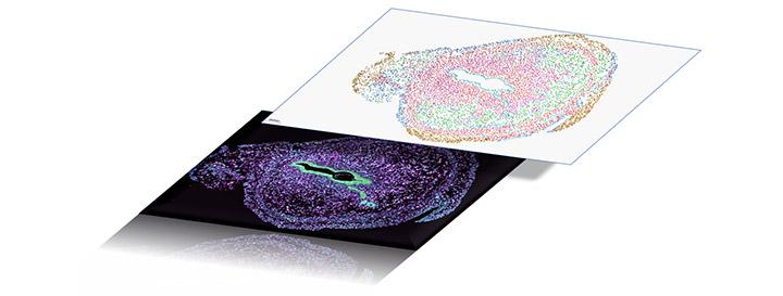
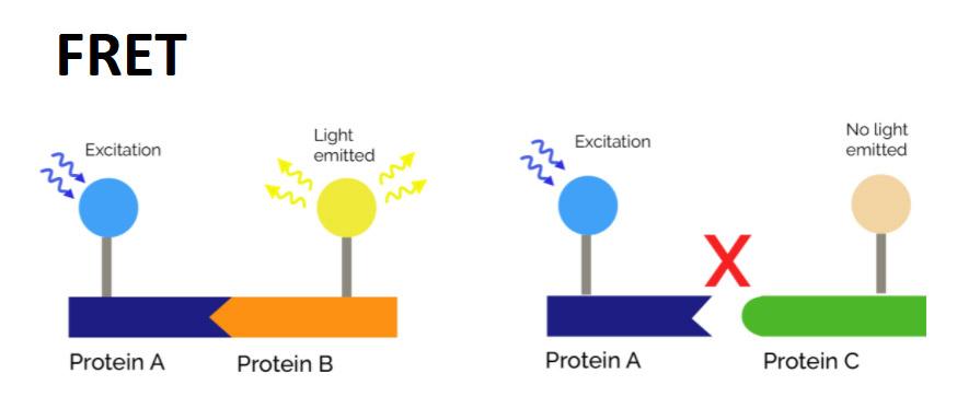
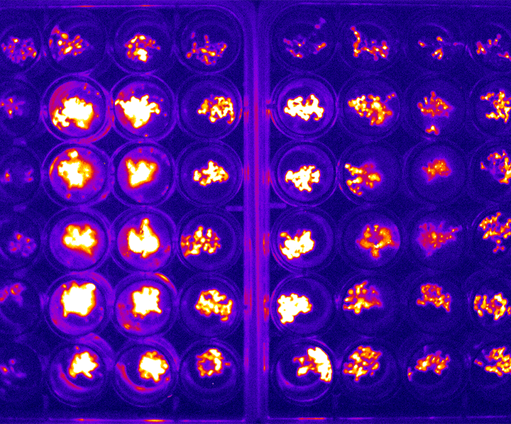
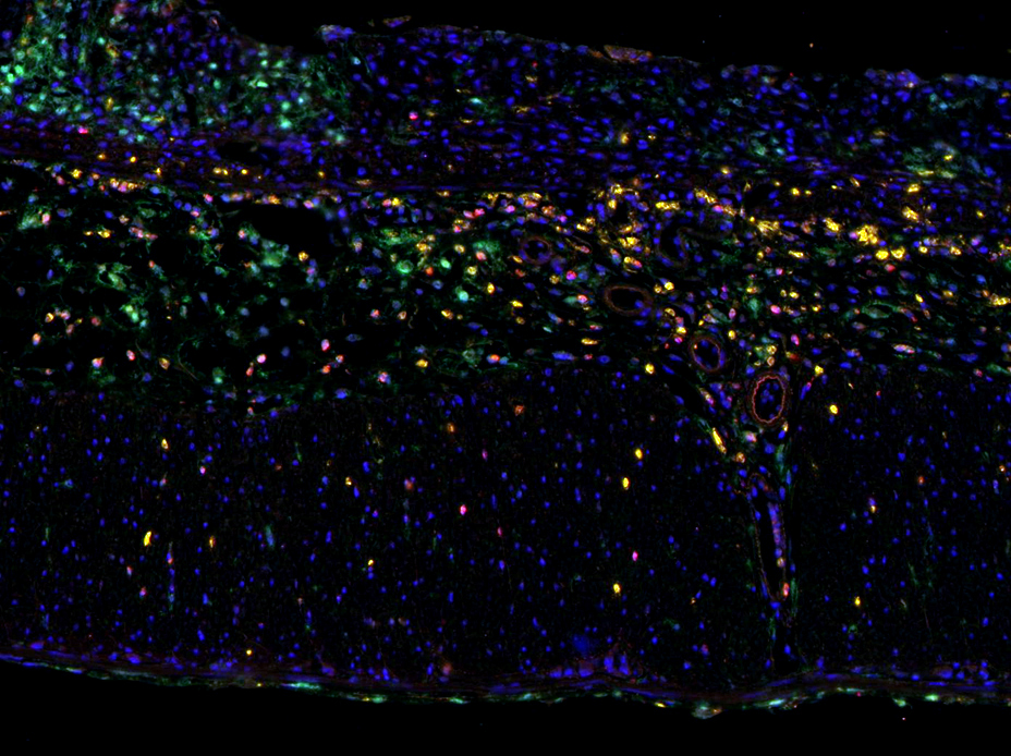
Andor offers a full range solutions for developmental biology: from sample preparation to image acquisition and analysis. For tissue sample preparation, the cryostats allow precise sectioning of the sample. Complete imaging solutions as the compact and affordable Andor Benchtop Confocal and the high-end imaging solution, the Andor Dragonfly. Highly sensitive EMCCD cameras to image low light and challenging samples, and sCMOS detectors to image high-speed and with high resolution. The workflow ends with image analysis options for cell biologists brought by different imaris packages: Imaris for Cell Biologists,

Learning Resources

Customer Publications

Author Title Journal Year
Kynan T Lawlor et al Cellular extrusion bioprinting improves kidneyorganoid reproducibility and conformation Nature Materials 2021
Bei Liu et al Chemically defined and xeno-free culture conditionfor human extended pluripotent stem cells Nature Communications 2021
Yan Zhang et al Oocyte-derived microvilli control female fertility byoptimizing ovarian follicle selection in mice Nature Communications 2021
Jifeng Liu et al Synthetic extracellular matrices with tailoredadhesiveness and degradability support lumenformation during angiogenic sprouting Nature Communications 2021
Anthony M Pettinato et al Sarcomere function activates a p53-dependent DNAdamage response that promotes polyploidizationand limits in vivo cell engraftment Cell Reports 2021
Julien Rességuier et al Organoid reproducibility and conformation Journal of Controlled Release 2021
Lama Tarayrah-Ibraheim et al DNase II mediates a parthanatos-likedevelopmental cell death pathway in Drosophilaprimordial germ cells Nature Communications 2021
Anthony M. Pettinato et al Development of a Cardiac Sarcomere Functional Genomics Platform toEnable Scalable Interrogation of Human TNNT2 Variants Circulation 2020
Yue Li Melatonin promotes human oocyte maturation and early embryodevelopment by enhancing clathrin‐mediated endocytosis J Pineal Res. 2019
Celine J. Vivien et al Vegfc/d-dependent regulation of the lymphatic vasculatureduring cardiac regeneration is influenced by injury context npj Regenerative Medicine 2019
Leily Kashkooli et al Ectoderm to mesoderm transition by down regulation ofactomyosin contractility Plos Biology 2019
Lorna J. Hale 3D organoid-derived human glomeruli for personalised podocyte disease modelling and drug screening Nature Communications 2018