
Applications
 Part of the Oxford Instruments Group
Expand
Collapse
Part of the Oxford Instruments Group
Case Studies
Author: Maria Alieva, Hubrecht Institute and Princess Máxima Center for Pediatric Oncology, and colleagues
Published: 01 Jan 2021 · Last updated: 06 Sep 2021
Tags: imaris-for-cancer-research-ar, cancer-research-lr
Researchers led by Maria Alieva from the Hubrecht Institute and Princess Máxima Center for Pediatric Oncology, both in the Netherlands, have incorporated Imaris software into an in vivo imaging approach they developed to study glioblastoma, a highly aggressive form of brain cancer.
Although tumor cell invasion is thought to be an important early event in glioblastoma’s progression, it has proven to be a difficult process to image. The new study provides a more detailed view of glioma invasion patterns, providing information that could help improve accuracy of prognosis and serve as a basis for personalized therapeutic approaches.
Previous studies performed using histological sections have shown that glioblastoma borders follow different invasion patterns. To find out how these patterns might be clinically relevant, Alieva and colleagues used time-lapse intravital imaging to study cancer cells that were implanted into the brains of mice.
“We acquired 3D time-lapse movies that showed tumor cells at the single cell level as well as other features from the tumor microenvironment such as the blood vessels,” said Alieva. “We used Imaris to track the behavior single cancer cells in 3D.”
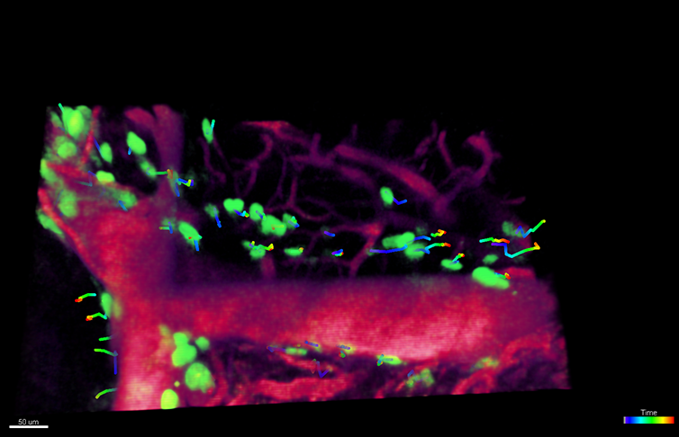
Researchers investigated the behavior of tumor cells (green) moving along blood vessels (red) or in the parenchymal space by tracking tumor cell movement over time with the Spot module of Imaris. Image courtesy of Maria Alieva, Hubrecht Institute and Princess Máxima Center for Pediatric Oncology.
The researchers used the Imaris Spot module to correct for any x-y drift that occurred over time by tracking reference objects that were static and then applying drift correction with rotational and translational registration. They also identified regions of interest in which to study tumor cell behavior and cropped them, since it is easier to work with smaller files.
“With the Spots module, we semi-automatically tracked single tumor cells over time,” said Alieva. “This allowed us to extract different features that described the behavior of single cells, such as position, speed, and persistence. Creating an object at the position of the tumor core also let us tell whether cells were moving towards or away from the tumor core.”
The researchers carried out imaging analysis on various tumor border patterns, including the invasive margin border pattern, diffuse infiltration border pattern, and well-defined tumor border pattern. This work represents the first time that distinct tumor border patterns found on histological sections have been correlated with specific tumor cell dynamics in vivo.
They found that although tumor cells were motile throughout the entire tumor including its core, invasive and diffuse margins contributed to tumor spreading in the brain. The invasive margin showed slow but directed invasion while the diffuse infiltration margin was characterized by fast, but less directed, movement.
The researchers are now investigating how environmental factors influence the behavior of diffuse intrinsic pontine glioma, a type of malignant brain tumor that affects children. Using different strategies to identify factors present in the tumor microenvironment, they want to better understand how these factors influence malignant tumor behavior.

© Oxford Instruments 2026
