
Applications
 Part of the Oxford Instruments Group
Expand
Collapse
Part of the Oxford Instruments Group

The Imaris for Neuroscientists package enables 3D reconstruction of neurons and arborization analysis. It can resolve various structures, such as axons and dendrites, somas, dendritic spines, microglia, or astrocytes. It calculates a range of neuron-specific measurements, such as dendrite or segment length, orientation, diameter, branch level, spine density and spine shape analysis and classification.
Free Trial Request Pricing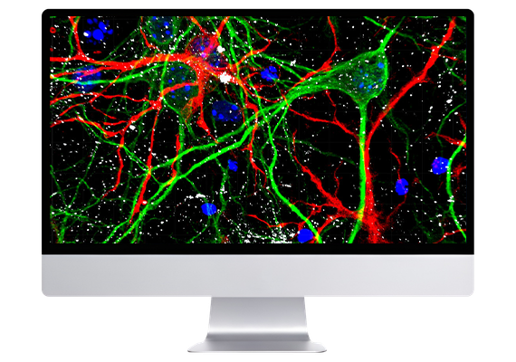
Imaris enables neuroscientists to visualise and analyse complex neural imaging datasets within a unified software environment. It supports reconstruction and quantification of neuronal structures, helping researchers interpret connectivity and organisation in both static and dynamic systems. The platform integrates tools for segmentation, tracking, and measurement, reducing reliance on multiple software packages. It is designed to handle large volumetric and time-resolved datasets generated by modern imaging technologies. Flexible workflows allow adaptation to diverse experimental designs, from single-cell analysis to large-scale brain imaging.
Microscopy image analysis requires precise detection of multiple biological structures based on the initial signal saved as an image data. Object detection in Imaris will be quick and easy as users are guided within a wizard driven interface to create perfect models of their data. With our patented Torch™ tool and several performance improvements tracing neurons with Autopath or Autodepth in dense and thick samples is very efficient and becomes a unique experience.
Imaris for Neuroscientists offers:
Glia cell structure and shape is related to their function and ability to interact with neuronal cells. Dysfunctions of glial cells can be associated with several neurodegenerative diseases. Neuroscientists study glia inside the brain tissue sections from both healthy animals and from transgenic mouse disease models. Due to the complexity of the neuronal glia images, 3D image analysis software is essential for acquiring the quantitative information and comparisons between samples. Imaris for Neuroscientists offers:
Automatic neuron tracing combines the benefits of an intensity-based method with a unique machine learning approach, providing the most flexible tool on the market for tracing a multitude of neuron types. Creating filament models is very easy, even for non-experienced users, because they are guided via precisely designed wizards with the result previewed after each step. By following the wizard instructions accompanied with a few paintbrushes along the structures of interest, they are separated from the background. Tracing neurons inside Imaris for Neuroscientists is:
Regardless of which filament tracing method is chosen (wizard driven automatic tracing or an Autopath/Autodepth mode) dendritic spines can be automatically detected and precisely modelled based on the threshold. The benefits of the automated dendritic spine detection method in Imaris are:
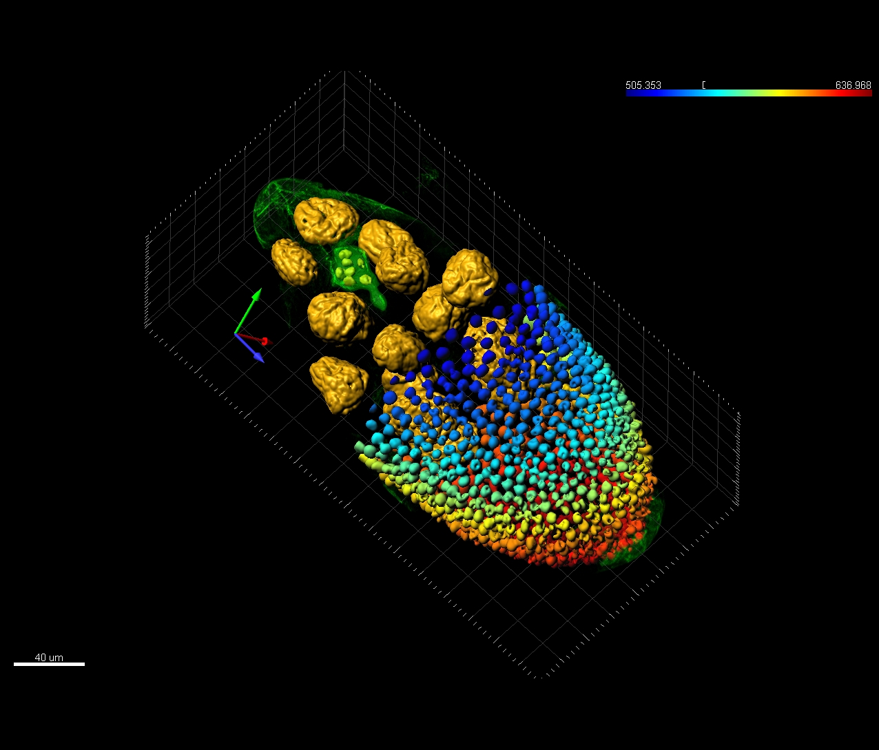
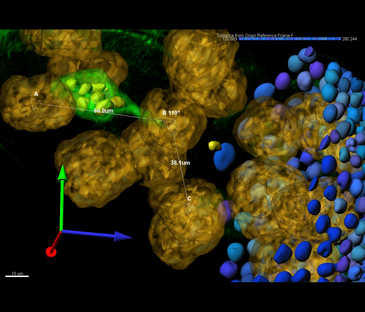
Surface objects define the boundaries of structures with varying shapes and sizes, such as cells, nuclei, and brain regions, while Spots model point-like or vesicle-like features, enabling rapid detection of thousands of structures. Together, Surface and Spots complement neuron tracing by providing additional measurements, including volume, position, and intensity. Imaris for Neuroscientists also enables automated distance measurements between detected structures, including filamentous and stained objects. In Imaris for Neuroscientists you can:
With Imaris 11, you can effortlessly create a customized image analysis protocol (Workflow) tailored to your specific research area and requirements. These Workflows can be saved and applied to other datasets with just one click, streamlining your analysis process. Discover how Workflows can accelerate your journey from microscopy data to meaningful insights, impactful publications, and engaging conference presentations.
The software supports a range of imaging workflows, from exploratory visualisation to advanced quantification and tracking. Its modular structure allows users to apply relevant tools depending on their experimental needs.

Imaris provides tools for tracing dendrites and axons within 3D datasets. This enables reconstruction of complex neuronal morphologies and supports detailed structural analysis. It is particularly valuable for studying connectivity and branching patterns.

The software enables identification and analysis of synaptic structures in dense datasets. Users can quantify spatial distribution and relationships between synapses and neurons. This supports deeper understanding of neural connectivity.

Imaris supports tracking of dynamic processes such as neurite growth and cell movement over time. This allows researchers to study development and response to stimuli. Tracking tools help quantify changes in neuronal behaviour.
Cell Biology is areas of research in life sciences that focus on the fundamental processes of life. Cell biology encompasses a broad range of research areas and applications such as apoptosis, cell cycle & cell division, DNA damage, plant cell biology, vesicle trafficking, in vitro studies etc. As for model organisms, cell biology investigates them all, from the most simple prokaryotes (bacteria) to single-cell eukaryotes (yeast, fungus) and even multicellular organisms.
Basic cancer research combines several aspects of studying cancer cell phenotypes, gene expression and interactions with the microenvironment in vitro and in vivo to better understand carcinogenesis, malignancy and develop new potential therapies. Cancer research often requires applying advanced fluorescence microscopy to study cancer cell behaviour interaction with the environment and spatial distribution of the tumour models in a time lapse.
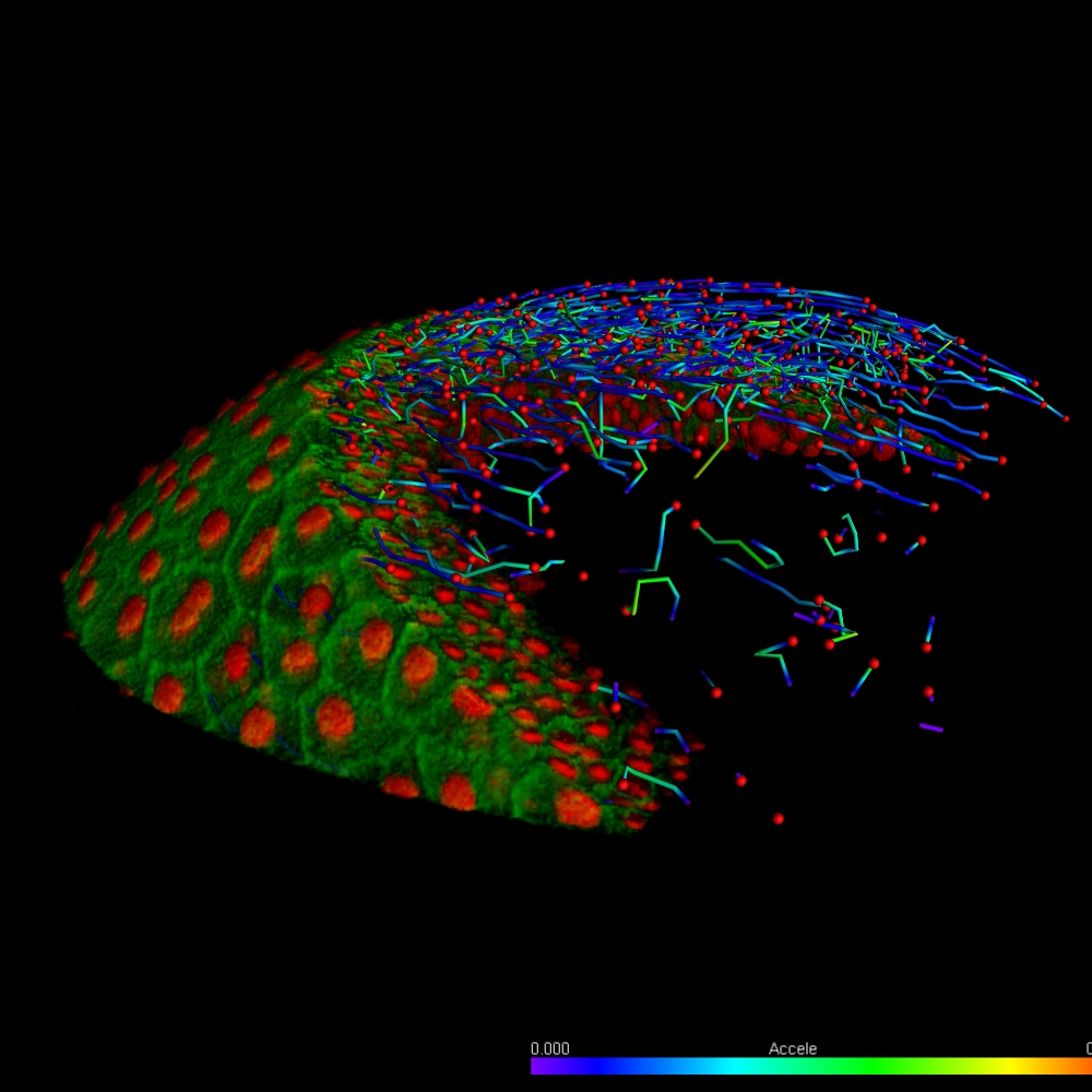
Developmental Biology are areas of research in life sciences that focus on the fundamental processes of life. Developmental biology studies the process by which multicellular organisms grow and develop. Research focuses on processes such: as metamorphosis, embryonic development, tissue growth, morphogenesis, stem cell differentiation, embryogenesis, plant development and regeneration. Research in these areas is done both on a microscopic and molecular level, and multiple technologies are needed to successfully accomplish this work.
Neuroscience is a multidisciplinary branch of science focused on the study of the nervous system and how the brain works. The field studies nervous system functions, brain function and the related structures such as the spinal cord. It combines anatomy, physiology, cytology, molecular biology, developmental biology and modelling in order to understand neurons and neuronal circuits. As neuroscientists often balance on the cutting edge of science, they require sophisticated methods such as fluorescence labelling, optogenetics, photostimulation and state of the art image analysis.
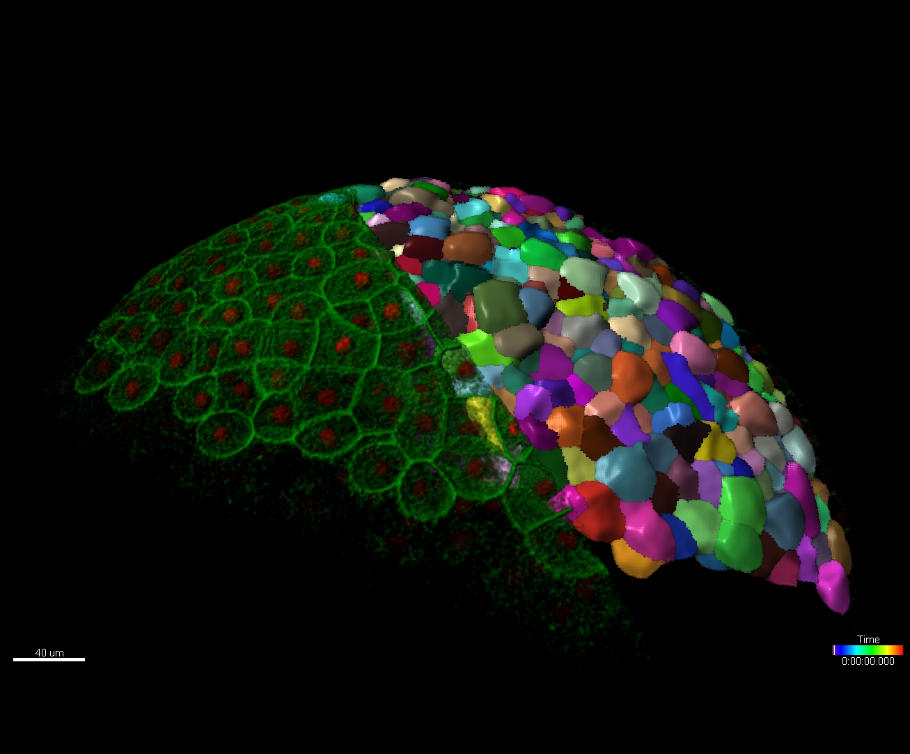
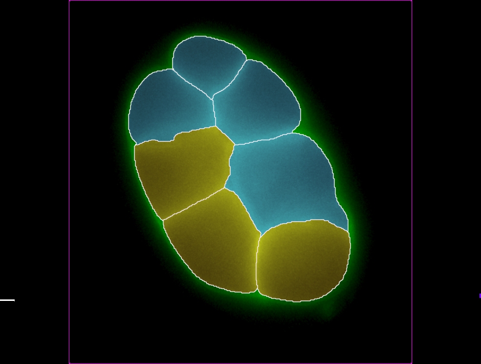
Automated classification of detected objects (cells, nuclei, vescicles) with a trainable Machine Learning Classifier (ML), based on selected statistic or the combination of 2 features. Classes are labelled and available for visual presentation, plotting and for downstream analysis (export of statistics).

© Oxford Instruments 2026
 Powered by Bioz