
Applications
 Part of the Oxford Instruments Group
Expand
Collapse
Part of the Oxford Instruments Group

Imaris for Tracking provides interactive processing, visualization and analysis software for 3D and 4D microscopic images. Featuring state of the art volume rendering, plus object detection and tracking tools Imaris for Tracking is perfect for researchers who want to automatically analyze moving objects including those that divide over time, create a lineage tree plot and generate quantitative information from their image data.
Free Trial Request Pricing
Imaris provides an integrated environment for tracking dynamic biological processes within multidimensional microscopy data. It supports automated and semi-automated workflows that reduce manual tracking effort and improve reproducibility. By combining tracking with segmentation and quantification tools, users can extract detailed motion and interaction data from complex datasets. The platform handles large time-series datasets, making it suitable for modern imaging workflows.
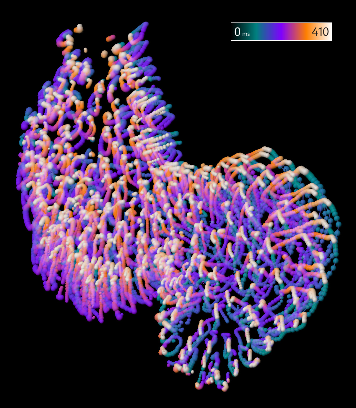
Imaris for Tracking gives the choice of proven Imaris 3D tracking algorithms: Brownian Motion, Autoregresive Motion (Expert), Connected Components or Lineage to generate the most accurate tracking results for different types of moving objects. Users can automatically correct translational and rotational drift or track within the new freehand coordinate system (Reference Frame) or use the automated option for drift correction.
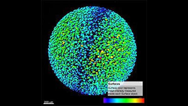
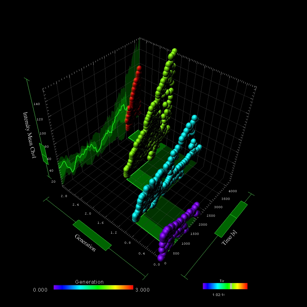
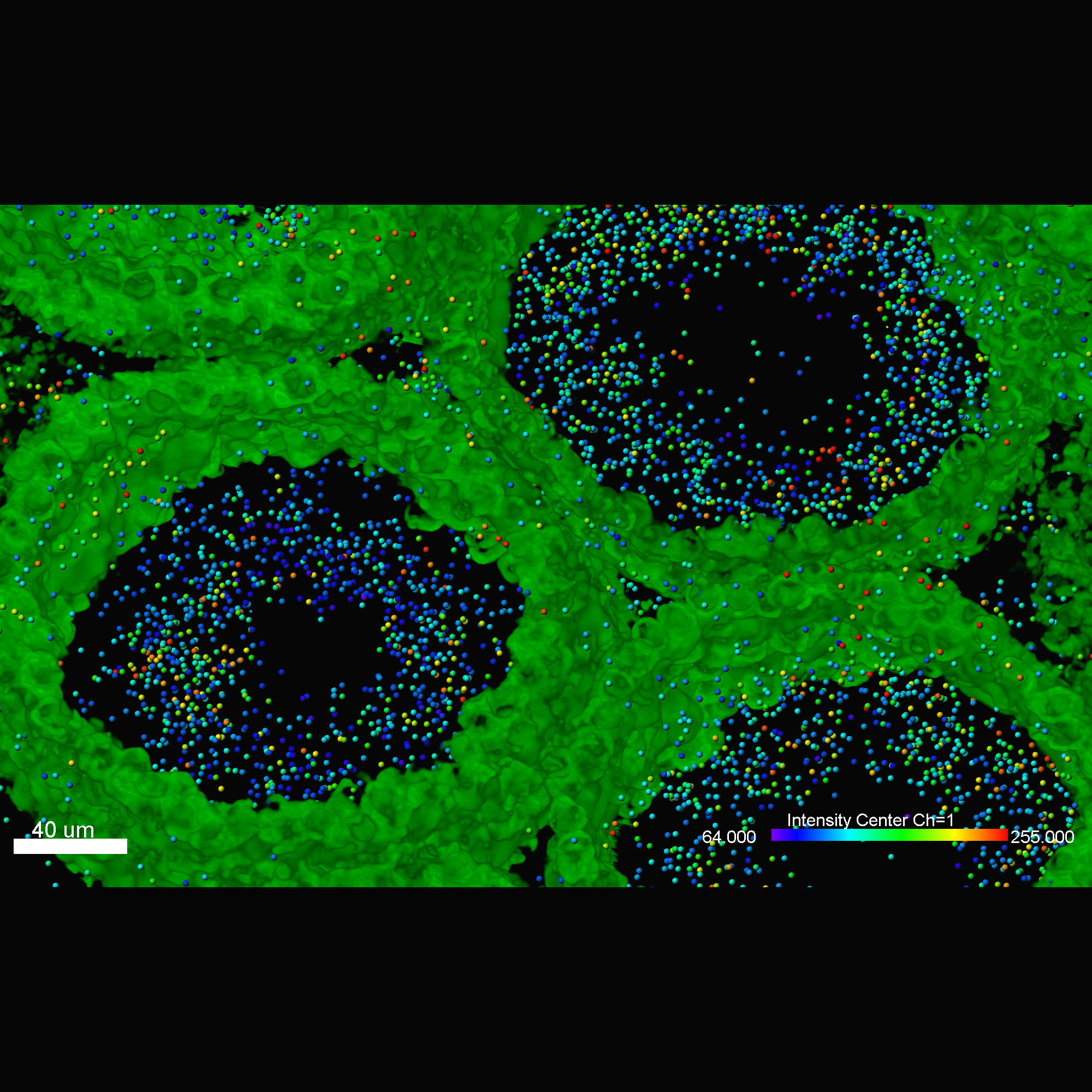
After segmenting the image data Imaris calculates a wide range of statistics through time for all detected objects, Surfaces and Spots. All values can be used for color coding, plotted inside Imaris (using Vantage plots) or exported in an .csv or .xls file format. Parameters presented below are the most common statistic types needed by biologists. Imaris reports many more.
Imaris for Tracking enables easy manual editing of the results of automated tracking. Use Labels to add additional descriptive parameters.
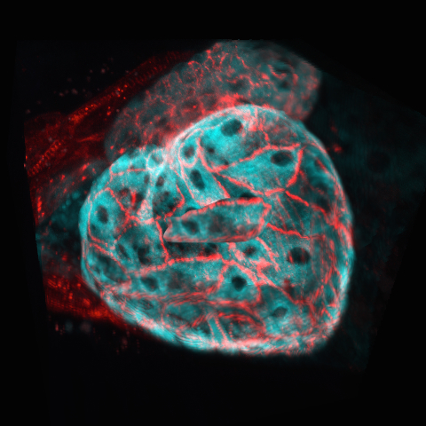
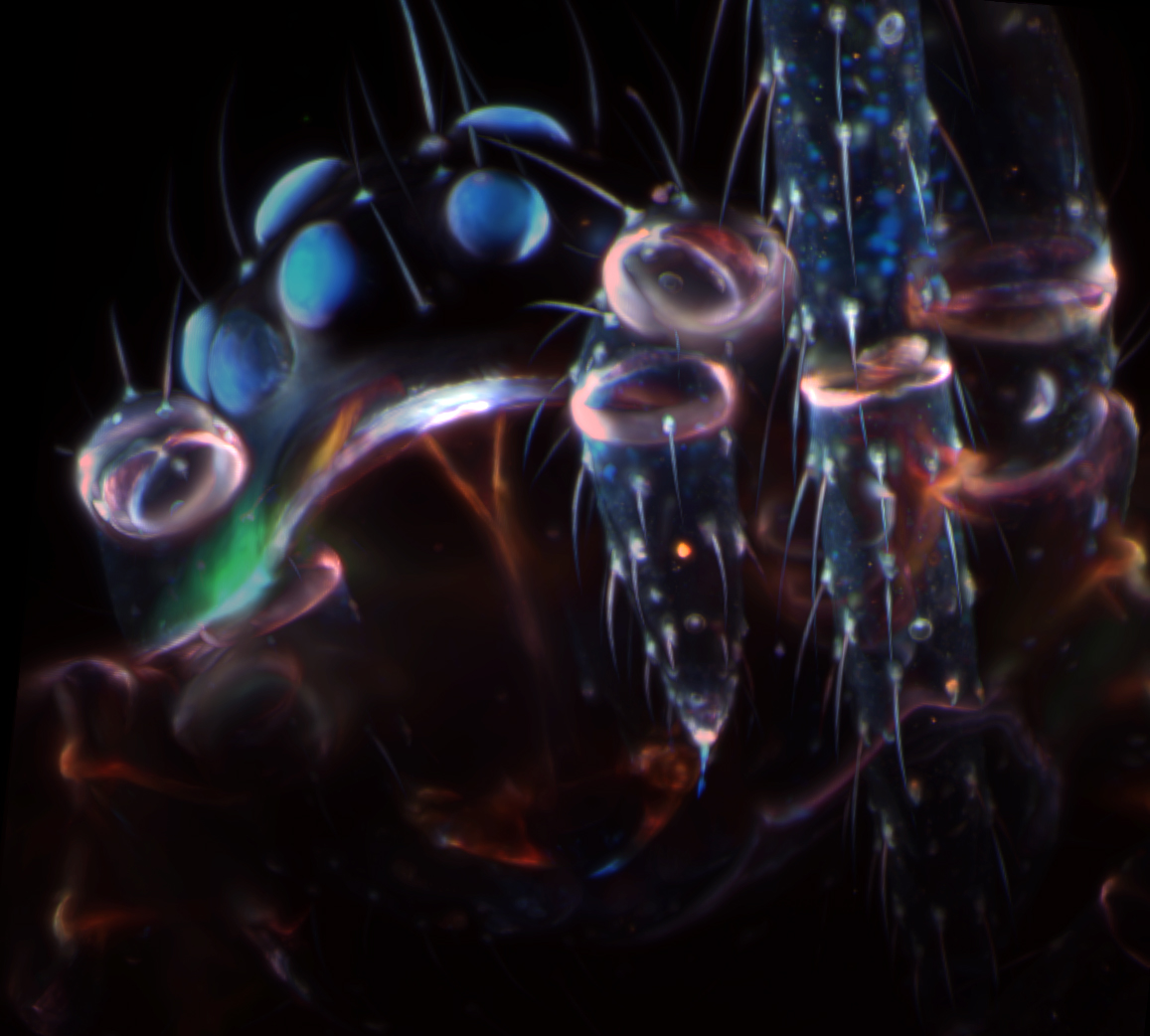
Imaris for Tracking provides a complete set of features for visualization of multi-channel microscopy datasets from static 2D images to 3D time series regardless of their size and format and multiple image and video export options to enhance your papers and presentations.
With Imaris 11, you can effortlessly create a customized image analysis protocol (Workflow) tailored to your specific research area and requirements. These Workflows can be saved and applied to other datasets with just one click, streamlining your analysis process. Discover how Workflows can accelerate your journey from microscopy data to meaningful insights, impactful publications, and engaging conference presentations.
The software supports a range of imaging workflows, from exploratory visualisation to advanced quantification and tracking. Its modular structure allows users to apply relevant tools depending on their experimental needs.

Imaris for Tracking uses automated algorithms to identify and follow objects across time-lapse datasets. This reduces the need for manual tracking while maintaining consistency across large experiments. It is particularly useful for analysing complex motion patterns in dense or dynamic samples.

Users can manually review and refine tracking results within the same environment. Tracks can be corrected, split, or merged with immediate visual feedback, helping ensure accurate datasets. This combination of automation and control supports reliable analysis in challenging imaging conditions.

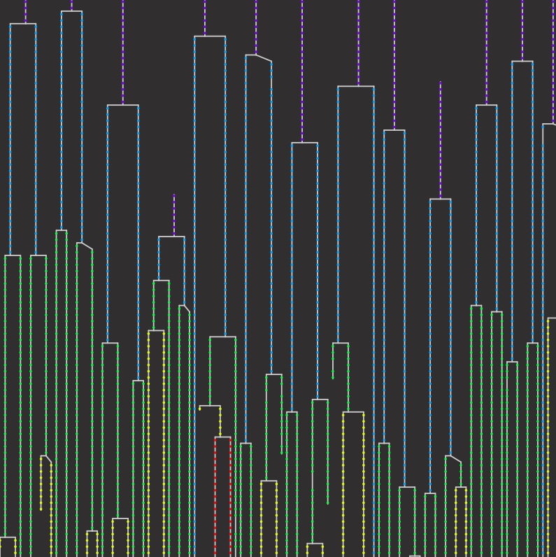
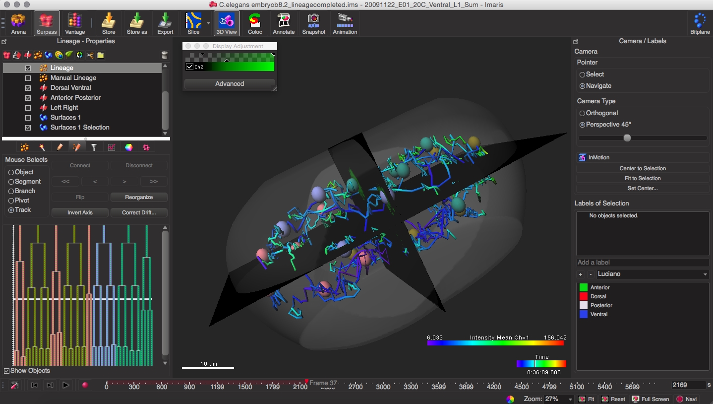
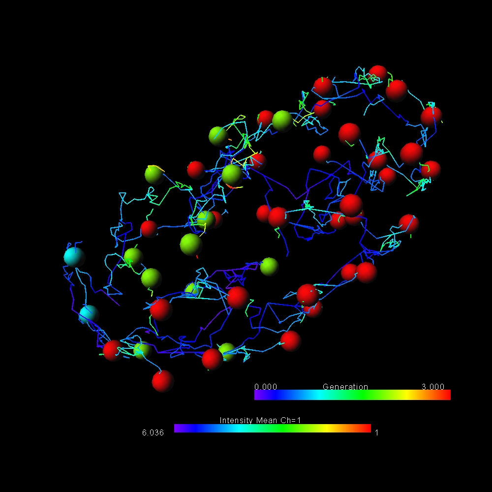
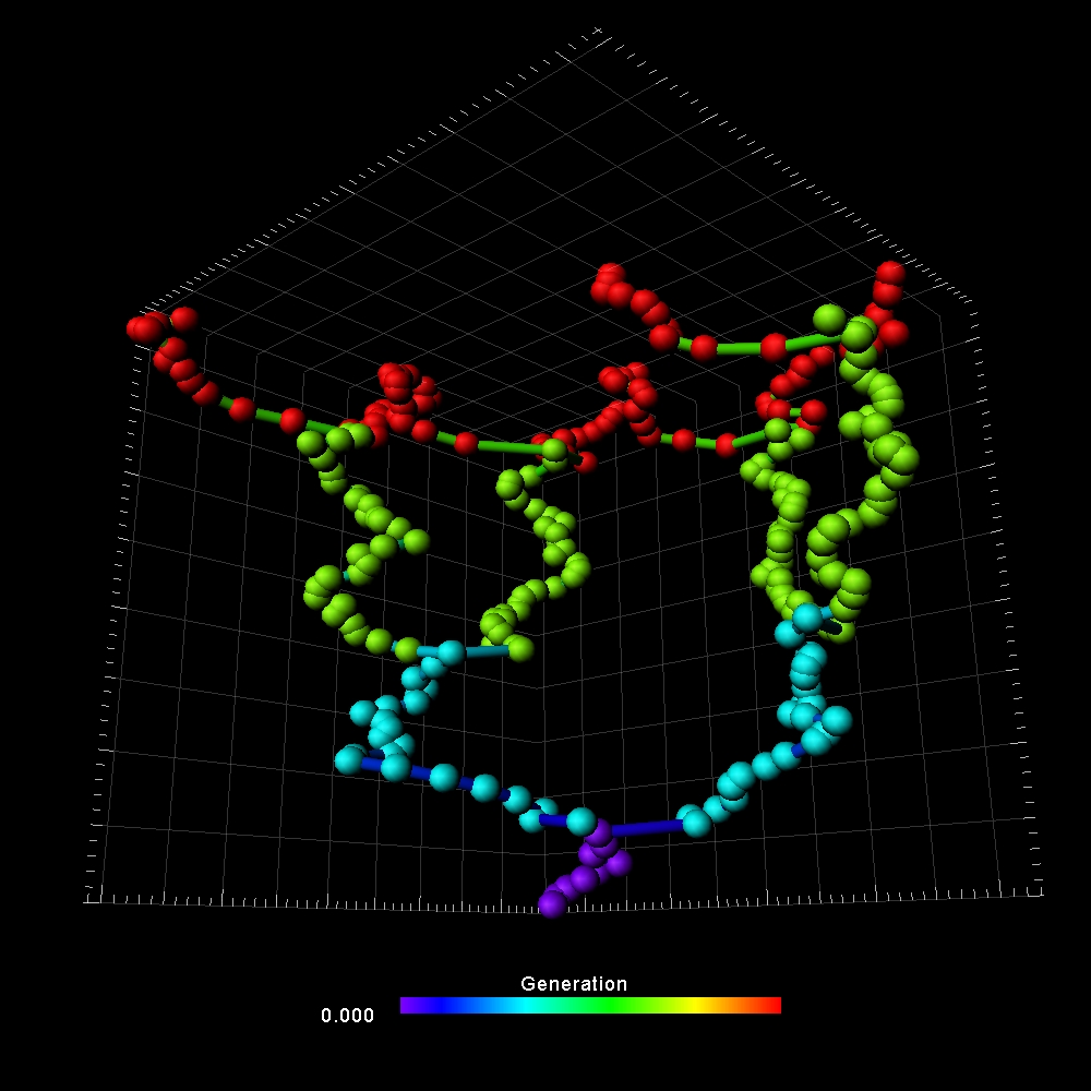
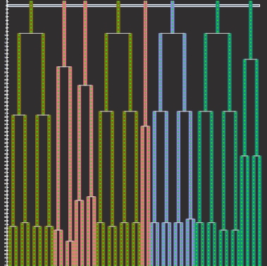
Imaris supports tracking of object division and lineage relationships over time. This enables users to follow how cells split and evolve, creating hierarchical track structures. It is especially valuable in studies of development, proliferation, and cell behaviour over extended time series.
Cell Biology is areas of research in life sciences that focus on the fundamental processes of life. Cell biology encompasses a broad range of research areas and applications such as apoptosis, cell cycle & cell division, DNA damage, plant cell biology, vesicle trafficking, in vitro studies etc. As for model organisms, cell biology investigates them all, from the most simple prokaryotes (bacteria) to single-cell eukaryotes (yeast, fungus) and even multicellular organisms.
Basic cancer research combines several aspects of studying cancer cell phenotypes, gene expression and interactions with the microenvironment in vitro and in vivo to better understand carcinogenesis, malignancy and develop new potential therapies. Cancer research often requires applying advanced fluorescence microscopy to study cancer cell behaviour interaction with the environment and spatial distribution of the tumour models in a time lapse.
Developmental Biology are areas of research in life sciences that focus on the fundamental processes of life. Developmental biology studies the process by which multicellular organisms grow and develop. Research focuses on processes such: as metamorphosis, embryonic development, tissue growth, morphogenesis, stem cell differentiation, embryogenesis, plant development and regeneration. Research in these areas is done both on a microscopic and molecular level, and multiple technologies are needed to successfully accomplish this work.
Neuroscience is a multidisciplinary branch of science focused on the study of the nervous system and how the brain works. The field studies nervous system functions, brain function and the related structures such as the spinal cord. It combines anatomy, physiology, cytology, molecular biology, developmental biology and modelling in order to understand neurons and neuronal circuits. As neuroscientists often balance on the cutting edge of science, they require sophisticated methods such as fluorescence labelling, optogenetics, photostimulation and state of the art image analysis.
Automated classification of detected objects (cells, nuclei, vescicles) with a trainable Machine Learning Classifier (ML), based on selected statistic or the combination of 2 features. Classes are labelled and available for visual presentation, plotting and for downstream analysis (export of statistics).

© Oxford Instruments 2026
 Powered by Bioz