
Applications
 Part of the Oxford Instruments Group
Expand
Collapse
Part of the Oxford Instruments Group

Imaris is an advanced 3D and 4D image visualisation, analysis and interpretation software platform designed for microscopy data. It enables researchers to explore complex biological structures and dynamics from large, multidimensional datasets. Widely used in life sciences, Imaris supports workflows such as cell tracking, segmentation and quantitative analysis. Its intuitive interface helps users move from raw image data to meaningful insights efficiently. It is commonly applied in areas such as neuroscience, cell biology and developmental biology.
Free Trial Request PricingImaris provides a comprehensive environment for managing, visualising and analysing high-content microscopy data. It integrates powerful rendering with quantification tools, enabling users to extract biologically relevant information from large datasets. The platform is designed to handle increasing data complexity while maintaining usability for both experienced and non-specialist users. Its modular approach supports tailored workflows, from simple visualisation to advanced analysis. Automation and reproducibility are emphasised to support reliable research outcomes. Integration with existing imaging systems and data formats supports broad adoption across laboratories.
At certain stages of searching for and evaluating new cancer therapies, whether it’s a chemotherapy, immunotherapy or radiotherapy, cell cultures or spheroids, researchers pick fluorescence microscopy to visualize the subtle and dynamic interaction and changes in the system. Imaris can directly open and visualize microscopy datasets in all formats available in the market. Imaris offers unique segmentation/object detection tools (like Spots and Surfaces) to better visualize and understand the sample in 3D.
Imaris for Cell Biologists is the most powerful commercial software for live cell tracking & analysis. It rises to the challenge of monitoring temporal changes in biological systems. Users have a choice of multiple sophisticated automatic tracking algorithms, manual tracking options as well as an intuitive and interactive track editing tool.
Deconvolution is often a first and important step in image analysis when users need to sharpen their images. Imaris overcomes common workflow problems where users need to open their files in various software packages, convert images and save multiple copies, by integrating deconvolution functionality into the Imaris menu. The main advantages of Imaris ClearView™ deconvolution are:
Microscopy image analysis requires precise detection of multiple biological structures based on the initial signal saved as an image data. Object detection in Imaris will be quick and easy as users are guided within a wizard driven interface to create perfect models of their data. With our patented Torch™ tool and several performance improvements tracing neurons with Autopath or Autodepth in dense and thick samples is very efficient and becomes a unique experience.
Analysis of the neuronal branches and their structure is a valid neuroscience research tool and can be accelerated with a dedicated 3D image analysis software. Imaris for Neuroscientists offers:
Imaris for Tracking gives the choice of proven Imaris 3D tracking algorithms: Brownian Motion, Autoregresive Motion (Expert), Connected Components or Lineage to generate the most accurate tracking results for different types of moving objects. Users can automatically correct translational and rotational drift or track within the new freehand coordinate system (Reference Frame) or use the automated option for drift correction.
With Imaris 11, you can effortlessly create a customized image analysis protocol (Workflow) tailored to your specific research area and requirements. These Workflows can be saved and applied to other datasets with just one click, streamlining your analysis process. Discover how Workflows can accelerate your journey from microscopy data to meaningful insights, impactful publications, and engaging conference presentations.
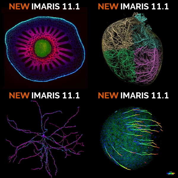
Analyzing heart vasculature is challenging: complex vessel networks require precise segmentation and comparing cardiac compartments demands consistent methodology. Imaris 11 Workflows integrate machine learning pixel classification for automated vessel segmentation with Filament Tracer for precise quantification of vessel length and diameter, enabling reproducible analysis across multiple heart samples and compartments.
= Optional
Automated classification of detected objects (cells, nuclei, vescicles) with a trainable Machine Learning Classifier (ML), based on selected statistic or the combination of 2 features. Classes are labelled and available for visual presentation, plotting and for downstream analysis (export of statistics).
Imaris supports 3D and 4D microscopy datasets from techniques such as confocal and light sheet imaging. Supported file formats and data sizes depend on configuration.
The platform is designed to handle large volumetric datasets efficiently. Performance depends on system hardware and dataset size.
Imaris supports segmentation, tracking and quantitative measurement workflows. Additional capabilities may be available through optional modules.

© Oxford Instruments 2026
 Powered by Bioz