
Applications
 Part of the Oxford Instruments Group
Expand
Collapse
Part of the Oxford Instruments Group
Case Studies
Published: 10 Feb 2021 · Last updated: 18 Oct 2023
Stem Cell Biology
Dr. Alex Y. Huang’s research team from Case Western Reserve University in Cleveland, Ohio, has used Imaris to observe how human mesenchymal stem cells (hMSCs) behave in response to an inflammatory challenge. Understanding the in vivo mode of action for these cells could help scientists improve their clinical efficacy in aiding bone repair and wound healing and as therapy for diseases and conditions such as Parkinson’s disease, Crohn’s disease, and diabetes.
Human mesenchymal stem cells are being studied as possible therapies for a wide variety of diseases because of their ability to modulate the immune system, differentiate into various cell types, and to accumulate at disease sites. However, scientists don’t completely understand how hMSCs exert their therapeutic effects and immune modulation in vivo. Granulocytes are a key part of the innate immune system, and Dr. Huang points out that how granulocytes in bone marrow respond to invading pathogens has not been well studied, especially at early time points in the minutes to hours range.
A high-resolution view
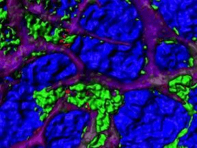
The researchers used intravital two-photon microscopy to gain a high-resolution spatial-temporal view of granulocytes in the bone marrow interacting with hMSCs injected into mice challenged with lipopolysaccharide, which evokes an inflammatory response.
“The Imaris software enabled us to measure granulocyte mobility, phagocytosis of cellular fragments, and interactions between granulocytes and the hMSCs from a dynamic 4-D imaging sequence,” Dr. Huang says.
The investigators analyzed the high-resolution fluorescent 4D imaging data with Imaris software, using the ImarisXT Channel Subtraction XTension to determine the localization of the hMSCs (labeled with Cell Tracker Orange) in relation to the granulocytes (labeled with GFP), in the bone marrow. Specifically, they used the Matlab Channel Arithmatics Function in ImarisXT to create a new channel in which the Cell Tracker Orange signals that overlapped the GFP were removed by subtraction.
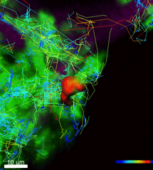
Cell tracking
The investigators also identified cells and traced tracked them using the Spots detection and tracking Analysis Function inalgorithms in Imaris. Finally, they exported the 3D spatial and time data for the hMSCs and granulocytes to Matlab for further analysis of how frequently these cells interacted and analyzed the direction that the granulocytes migrated in relation to the hMSCs.
“Spot tracking was helpful for evaluation of cell mobility and the channel subtractions were particularly useful in differentiating signal overlap, which allowed us to determine the intracellular uptake of hMSC cellular fragments by granulocytes,” Dr. Huang says.
The image analysis revealed that many of the hMSCs infused into the mice remained intact even though they repeatedly came into contact with host granulocytes. However, the researchers did observe granulocytes destroying some hMSCs, which was followed by phagocytosis and migration of cells containing hMSC fragments.
Research Paper: Jay T. Myers, Deborah S. Barkauskas, and Alex Y. Huang. Dynamic Imaging of Marrow-Resident Granulocytes Interacting with Human Mesenchymal Stem Cells upon Systemic Lipopolysaccharide Challenge. Stem Cells International, Vol. 2013, Article ID 656839, 11 pages, 2013. doi:10.1155/2013/656839.

© Oxford Instruments 2026
