Imaris software offers Biofilm researchers a powerful and flexible image visualization and analysis tool to enhance their research.
With Imaris you gain the ability to simply process and reconstruct data in 3D and fully automate segmentation and quantitative characterization of biofilm structures. Its flexibility allows all segmentation parameters to be set interactively in accordance with your scientific requirements.
“ The user-friendly interface of the Imaris software allowed us to easily process and re-construct the 3D images. ”
Dr. Jiunn Fong, University of California-Santa Cruz
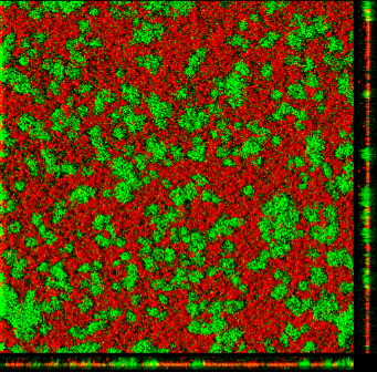
LEFT - This confocal laser scanning microscope image shows two strains of V. cholerae cultured together. One strain lacks VPS (green, GFP) and the other lacks matrix proteins (red, RFP).
Research Paper - Molecular architecture and assembly principles of Vibrio cholerae biofilms. Berk V, Fong JC, Dempsey GT, Develioglu ON, Zhuang X, Liphardt J, Yildiz FH, Chu S. Science. 2012 Jul 13;337(6091):236-9.
Other published biofilms papers using Imaris...
")
")
: p. 238–248.)") Bridier, A - Click to viewHong, S.H - Click to viewWang, R et al - Click to view
Bridier, A - Click to viewHong, S.H - Click to viewWang, R et al - Click to view
Discover how Imaris can assist your research...
| Biofilm characterisation |
Imaris feature |
| 1. Characterize the spatial and population heterogeneity within a biofilm |
✔ Create surface or spot objects based on colour and intensity |
| 2. Biovolume (μm3/μm2) calculated based on total volume (μm3) per area (μm2) |
✔ Measure the volume area of objects and images |
| 3. Biofilm compactness calculated based on total fluorescence per volume |
✔ Make use of voxel intensity statistics and object measurements |
| 4. Ratio between number of dead cells and number of living cells per volume |
✔ View the ratio between two different channels fluorescence |
CellPress webinar on "Formation and Dynamics of Biofilms"...
Hardly the completely solitary creatures they are often made out to be, microbes can form surface-associated communities, called biofilms, where they live and interact with each other within an extracellular matrix. Biofilms can have important roles in disease - for example, on indwelling medical devices where one feature of biofilm growth is increased resistance to antibiotics.

How biofilms assemble, mature, and evolve both in vitro and in vivo is the focus of the webinar that was presented by Cell Press on “Formation and Dynamics of Biofilms” in September 2013. We hope that you will enjoy hearing from Thomas Bjarnsholt, Joao Xavier, and Aaron Mitchell (left to right in the image) on the latest findings in the exciting and growing field of biofilm research.


")
")
: p. 238–248.)")