
Applications
 Part of the Oxford Instruments Group
Expand
Collapse
Part of the Oxford Instruments Group
Imaris has a proven track record as an enabling tool in the fields of Stem Cell and Regenerative Medicine - see examples below.
Imaris offers best in class 3D/4D visualization, automated & reproducible analysis and results exploration.
Researchers from the fields of Stem Cell and Regenerative Medicine often use Imaris' particle/cell tracking tools, its filament tracing and tracking features as well as the multi-dimensional data exploration plot generator (Vantage).
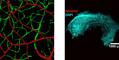
Studying vessel morphology and blood flow in the brain’s subependymal zone
James C. Culver and colleagues, Baylor College of Medicine
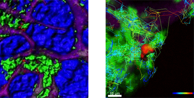
Dynamic imaging of bone marrow mesenchymal stems cells in response to immune challenge
Dr. Alex Y. Huang and colleagues, Case Western Reserve University
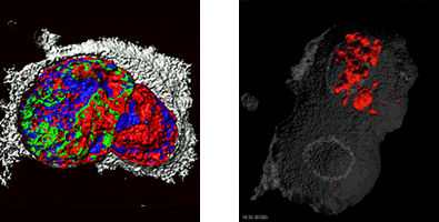
A 3-D look at cell division in cancer stem cells
Hong-Wu Xin and colleagues, National Cancer Institute, National Institutes of Health
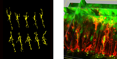
Studying dynamic stem cell interactions in the mouse neocortex
Dr. Branden R. Nelson and colleagues, University of Washington
| Feature of Stem Cells behaviour | Imaris feature |
|---|---|
| 1. Intra- and inter-cellular relationships during cell division and differentiation. | ✔ Use ImarisCell to detect cells and organelles. Automatically or interactively highlight sub populations based on one or more measured parameters. |
| 2. Cell-, organelle- or particle-motility analysis. | ✔ Use Surfaces, Spots or ImarisCell to detect the features of interest and then track their motion with ImarisTrack |
| 3. Find unique features of different cellular morphologies and compare them with each other | ✔ Use ImarisVantage to create explorative interactive plots. Any of the hundreds of calculated parameters can be used to build the plot (x,y,z, color and scale). |
| 4. Analyse relative location and movement of two types of cells in 3D time-lapse series. | ✔ Use Imaris XT functions to measure distance between objects in all time points. |
| 5. Observe nuclei and protein position hidden inside cell during the cell division. | ✔ Use a combined rendering approach (surface plus volume) along with a clipping plane or othoslicer to reveal hidden structures. |
| 6. Vasculogenesis, angiogenesis and Lymphangiogenesis. | ✔ Use FilamentTracer to detect blood and lymph vessel networks. Report segment length, diameter, tortuosity as well as network complexity. |
“ Being able to accurately reconstruct the fluorescence data into real three dimensional objects with unique parameters allows us to ask a much wider range of questions and get meaningful results. ”
Dr. Sansom, Beatson Institute of Cancer Research, Glasgow
 Direct In Vivo Evidence for Tumor Propagation by Glioblastoma Cancer Stem Cells. PLoS ONE 6(9): e24807. doi:10.1371/journal.pone.0024807")
 Evidence that Doublecortin Is Dispensable for the Development of Adult Born Neurons in Mice. PLoS ONE 8(5): e62693. doi:10.1371/journal.pone.0062693")
 HGF Expressing Stem Cells in Usual Interstitial Pneumonia Originate from the Bone Marrow and Are Antifibrotic. PLoS ONE 8(6): e65453. doi:10.1371/journal.pone.0065453")
Lathia JD - Click to viewMerz K et al - Click to viewGazdhar A et al - Click to view
ImarisCell features automated methods to identify, segment, trace, classify and analyze Cell, Nucleus and Vesicle objects in 2D and 3D time-resolved images. The Cell creation wizard has been specifically designed and structured to impose specific biological constraints relevant for tracking analysis. The first part of the Cell creation wizard detects, segments and classifies all pertinent cells and sub-cellular components. The next section of the Cell creation wizard provides the tools to track the newly created objects representing Cell, Nuclei or Vesicles.
Find out more by browsing our full range of stem cell related material located in our learning centre.
“ The Imaris software enabled us to measure granulocyte mobility, phagocytosis of cellular fragments, and interactions between granulocytes and the hMSCs from a dynamic 4-D imaging sequence. ”
Dr. Huang, Case Western Reserve University, Cleveland

© Oxford Instruments 2026
