

Imaris for Cell Biologists
View Product
 Part of the Oxford Instruments Group
Using Imaris image analysis software, Leslie Thompson and Isabella Inez Sanchez from the University of California-Irvine School of Medicine and colleagues discovered that Huntington’s disease shares neuropathological hallmarks with other neurodegenerative diseases, namely amyotrophic lateral sclerosis (ALS) and frontotemporal dementia. The findings could lead to new ways to treat Huntington’s disease, which causes a variety of movement, cognitive and psychiatric problems.
In ALS and frontotemporal dementia, RNA-protein structures called stress granules accumulate in the cytoplasm of neurons. The researchers recently found that a key effector of stress granule assembly, the Ras GTPase-activating protein-binding protein 1 (G3BP1), also accumulates in granule structures in the superior frontal cortex of Huntington’s disease patients and R6/2 mouse models of the disease.
Much of what is known about stress granule biology is based on in vitro experiments that use exogenous stressors to induce stress granules in cultured cells over short periods of time. “Our new work demonstrates that stress granule pathology can be detected in Huntington’s disease mouse and human brain tissues without applying exogenous stress,” said Sanchez. “We believe that stress granules resulting from chronic stress, such as those involved in neurodegenerative disease, are likely very different from stress granules that form in vitro.”
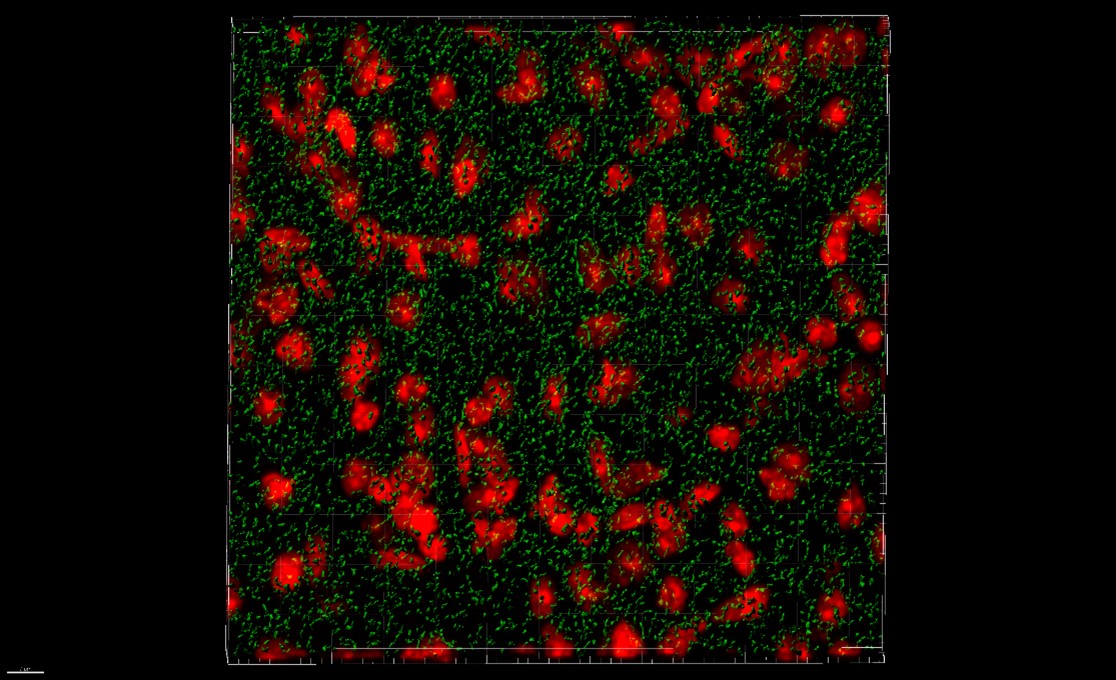
Surface rendering of G3BP1 granules (in green) detected by immunofluorescence in the Huntington's disease mouse model cortex. Image courtesy of Isabella Sanchez.
Stress granules that are induced in vitro have very discrete boundaries and are relatively easy to quantitate with 2-dimensional image processing software. “The moment we saw stress granules in brain tissue under the microscope, we realized they were not the same discrete punctate structures that assemble after inducing stress in cells in culture,” said Sanchez. “We needed to find a more stringent way to quantitate them.”
The researchers turned to the Imaris 3D surface rendering tool to acquire more information on stress granule count and volumes from Z-stack of images acquired with confocal microscopy. They did this by enabling the split touching objects function within the tool and using it to create masks of stress granules. They then normalized the stress granule count to the number of nuclei per frame, which was acquired using the Imaris Spots tool. After validating this process on induced stress granules in cultured cells, they applied the imaging pipeline to analyze stress granules in brain tissue.
The image analysis revealed an increase in stress granule density in brain tissue from a Huntington’s disease mouse model and human patients. The researchers also saw a reduction in stress granule density when cultured cells were treated with a microRNA that targets the stress granule nucleator G3BP1.
“Our findings suggest that testing small molecule compounds that modulate stress granule accumulation, such as microRNAs and other approaches, will be informative in future studies to evaluate the consequence of altered stress granule dynamics in Huntington’s disease,” said Thompson.
The researchers are now investigating a potential stress granule modulator in a mouse model of Huntington’s disease. “We plan to use the same Imaris image analysis pipeline to determine whether stress granule density or volume are affected in treated mice compared to controls,” said Thompson. “We hope that these studies help us assess whether targeting stress granule pathology is a viable therapeutic for the disease.”

© Oxford Instruments 2026