
Applications
 Part of the Oxford Instruments Group
Expand
Collapse
Part of the Oxford Instruments Group
In the central nervous system (CNS), microglia respond rapidly to tissue damage by extending cell processes and engulfing debris. Although scientists know that this process is important for brain development and CNS health, the mechanisms and kinetics of microglial phagocytic activity after CNS injury have not been well studied.
Researchers led by Geoffrey T. Norris from the University of Virginia are using Imaris to study phagocytosis of synaptic material by microglia. A better understanding of the biology involved in this process may help reveal the role that microglia phagocytosis plays in Alzheimer's disease or infections of the central nervous systems.
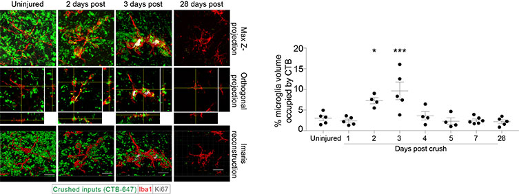
This figure details the kinetics of microglial engulfment with maximum phagocytic activity occurring on day three post-injury.
Quantifying engulfed neuronal debris
The researchers used acute CNS injury through optic nerve crush injury to study microglial phagocytosis mechanisms after injury in mice. Using Imaris combined with a protocol to assess interactions between CNS phagocytes and neurons, the researchers examined different time points during Wallerian degeneration. Wallerian degeneration, which occurs after a nerve fiber is crushed or severed distally from the neuronal cell body, is characterized by high levels of microglia phagocytosis of neuronal debris.
“The shapes and mask functions of Imaris allowed us to quantify engulfed neuronal debris within activated microglia,” said Norris. “The software’s intuitive user experience made this process much more effective.”
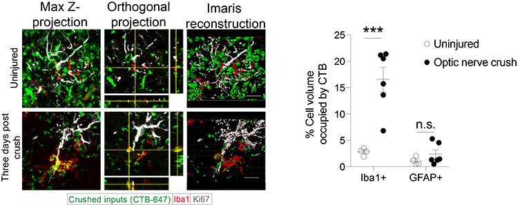
This figure details phagocytic activity of microglia (Iba1) and astrocytes (GFAP) on day three post injury when microglial phagocytosis is highest.
The researchers used Imaris to create 3D Surface and 3D volumetric renderings for microglia and engulfed debris. They then visualized and measured the debris volume by using the mask function of Imaris to remove any fluorescence from outside the microglia Surface. Next, they rendered the remaining engulfed fluorescence as a Surface and calculated the total volume of the engulfed debris. They determined the percentage of engulfment by dividing the volume of the internalized debris by the volume of microglial cells multiplied by 100.
Understanding debris clearance after injury
The image analysis allowed the researchers to discern that microglial phagocytic activity during CNS Wallerian degeneration occurs only on days two and three after injury and then drops sharply by day four post-injury. Additionally, like phagocytic processes that occur during development and other pathological processes, microglia make use of the complement system – a biochemical cascade of the immune system that helps to clear pathogens or debris, marking them for destruction. However, in contrast to processes that occur during development, neuronal activity is likely not a factor in driving microglial phagocytosis after injury.
“By understanding mechanisms of microglial phagocytosis, future work may be able to discern whether inhibiting or boosting microglial phagocytic activity may prevent or alleviate CNS dysfunction underlying human disease,” said Norris.
Next, the researchers plan to use Imaris to quantify phagocytic activity to determine which danger-associated molecular pattern molecules, or alarmins, are necessary for induction of microglial phagocytosis in acute injury to the brain and which receptors microglia use to recognize neuronal debris.

© Oxford Instruments 2026
