Daniel Düring and colleagues from University of Zürich/ETH Zürich in Switzerland and Max Planck Institute for Ornithology in Germany are combining expansion microscopy with conventional light sheet microscopy to make new discoveries about the brain of a small songbird known as the zebra finch. These birds are often used to study vocal learning, which can provide insights into human speech acquisition.
Expansion microscopy is a pre-processing method that uses a polymer system to physically expand a biological sample before imaging. The approach is typically used for cell cultures or thin brain slices, but in the new work, the researchers applied it to large portions of brain tissue by combining it with light-sheet microscopy.
The new approach, which the researchers call expansion microscopy and light sheet microscopy (ExLSM), enables imaging of larger volumes than have previously been possible with expansion microscopy while also producing images with higher resolution compared to conventional light sheet microscopy.
“ExLSM provides advantages in terms of imaging speed and photobleaching over the conventional approach of sectioning the brain for confocal microscopy, plus the added benefit of being able to image entire, unsectioned brain areas,” said Düring. “These benefits make it possible to study the underlying anatomy of song learning in great detail and much faster than previously possible.”
Zooming in on Song Nuclei
Düring’s research team is focused on resolving the network architecture of vocal learning and production in the zebra finch. Songbirds have a unique neural anatomy in which functionally and anatomically discrete areas called song nuclei form an interconnected network. Using ExLSM, the researchers were able to expand an entire song nucleus and image it with sub-cellular resolution.
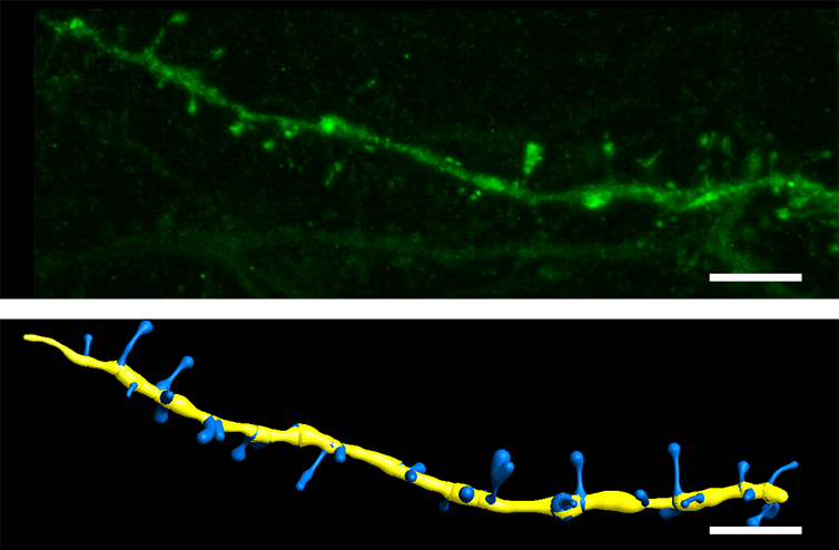
Researchers used Imaris to demonstrate the feasibility of ExLSM to resolve dendritic spine morphologies (top), which can be reconstructed easily with Imaris’ Filament Tracer tool (bottom). Reprinted under the Creative Commons Attribution License from Düring et al., 2019.
“Imaris provided a quick and easy way to evaluate the quality of our rather large volumetric datasets through maximum intensity projections,” said Düring. “The animation function of Imaris is a great tool to produce supplementing video sequences, which in this particular case, are much better suited to deliver an impression of a 3D dataset than 2D images.”
The researchers also used Imaris to demonstrate the feasibility of ExLSM to resolve dendritic spine morphologies, which can be reconstructed easily with Imaris’ filament tool. In unpublished analysis, they used the software’s spot tool to automatically extract neuron densities from their large datasets.
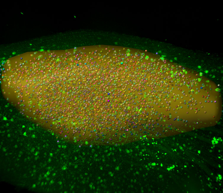
The researchers used Imaris’ Spots tool to automatically extract neuron densities from large datasets. The image shows how Imaris is used for automatic neuron counting in a large volumetric scan of a song nucleus acquired using expansion microscopy and light sheet imaging (ExLSM). Image courtesy of Daniel Düring.
Overall, the new study demonstrated the viability and advantages of using ExLSM for volumetric fluorescence imaging. The researchers are now using the approach to analyze the detailed structure of individual neurons in large-volume datasets. They hope this analysis will provide a new understanding of the structural variety within a given neuron population and the implications of this variety for the network architecture.
Research paper: Düring DN, Rocha MD, Dittrich F, Gahr M, Hahnloser RHR. Expansion Light Sheet Microscopy Resolves Subcellular Structures in Large Portions of the Songbird Brain. Front Neuroanat. 2019;13:2. Published 2019 Jan 31. doi:10.3389/fnana.2019.00002

