
Applications
 Part of the Oxford Instruments Group
Expand
Collapse
Part of the Oxford Instruments Group
Case Studies
Author: Dr. Branden R. Nelson and colleagues, University of Washington
Published: 10 Feb 2021 · Last updated: 18 Oct 2023
Stem Cells
As the nervous system develops, Notch signaling is crucial for maintaining the cells necessary to generate the correct numbers and types of neurons in different regions in the brain. Researchers led by Dr. Branden R. Nelson at the University of Washington in Seattle used Imaris software to better understand how cells interact to convey Notch signaling, which is dependent on cell contact.
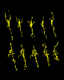
Isolated Dll1-active INP in the subventricular zone. Surface-rendered, segmented, and isolated INP in the subventricular zone that is active for a destabilized Dll1-d2YFP reporter. The Dll1-active INP extends and retracts multiple dynamic processes, including a long-range process back into the ventricular zone. Together with high-resolution static visualization of Dll1 protein immunolabeling, these high-resolution live cellular dynamics serve as the potential cellular basis for Dll1-mediated INP-to-RG feedback, contacting RG and driving higher levels of Notch signaling, helping to maintain their pool through development. Courtesy of Dr. Branden R. Nelson.
In the neocortex of mammals, radial glia stem cells generate cortical neurons by producing a transiently amplifying group of intermediate neurogenic progenitor cells. Higher levels of Notch signaling distinguish radial glia from intermediate neurogenic progenitors, which have lower levels of Notch signaling. Mutations or environmental factors that prevent or perturb normal progenitor interactions may play a role in human neurological disorders and disease.
“Although intermediate neurogenic progenitors have been shown to be a major molecular source for Delta-like 1 (Dll1)-mediated feedback to maintain higher Notch activity in radial glia, exactly how this feedback occurs was unknown,” explains Dr. Nelson. “Imaris proved crucial to our live imaging analysis and presentation of important radial glia and intermediate neurogenic progenitors cellular interactions.”
Cell-to-cell interaction
The researchers used confocal microscopy as well as high-resolution live-cell multiphoton microscopy to visualize fine process dynamics and cell-cell interactions in the intact niche. Using Imaris software, they conducted cell counts, process measurements, rotations, and manual reconstruction/segmentation of individual neocortical progenitors cells.
Dr. Nelson said that compared to other high-end software packages he has used, the learning curve for Imaris was much shorter because of its straightforward layout and simplified wizard and tutorial functions. He also liked the ease at which he could manipulate and crop individual 4D datasets, combine them into a single one, and, when necessary, correct for drift through the Fiji output function.
“It was very useful for this study to have the ability to easily surface render, segment, isolate, and psuedo-color different cellular morphologies using both Automatic and Manual Surface Functions in Imaris,” Dr. Nelson adds. “In fact, given the high density of labeling in some cases, the easy ability to manually draw contour lines for surface rendering was important for detailing interactions between different progenitor cell types.”
Overall, the image analysis revealed that at least some of the morphological stages of intermediate neurogenic progenitor cell migrations likely mediate the cellular basis for Dll1 feedback to radial glia. The investigators also uncovered how these diverse progenitors normally interact in their in situ neocortex stem cell niche, providing the basis for signaling that is dependent on cell-to-cell contact in different regions of the mammalian brain.
Two-color live-cell multiphoton orthoslice 4D view of the embryonic neocortical NSC niche reveals the "social network" of intermediate neurogenic progenitors [(INP) (green, transgenic Tbr2GFP)] and radial glia (RG)/INP (red, electroporated membrane-tdTomato) interactions. Imaris was used to combine multiple 4D datasets, correct for drift using Fiji output functions, and then allowed detailed sub-sampling of different regions in the 4D dataset at the single section level. Movie plays through the green channel first, then is repeated with both red and green channels. Courtesy of Dr. Branden R. Nelson.
Movie 2 and related figure. Isolated Dll1-active INP in the subventricular zone. Surface-rendered, segmented, and isolated INP in the subventricular zone that is active for a destabilized Dll1-d2YFP reporter. The Dll1-active INP extends and retracts multiple dynamic processes, including a long-range process back into the ventricular zone. Together with high-resolution static visualization of Dll1 protein immunolabeling, these high-resolution live cellular dynamics serve as the potential cellular basis for Dll1-mediated INP-to-RG feedback, contacting RG and driving higher levels of Notch signaling, helping to maintain their pool through development. Courtesy of Dr. Branden R. Nelson.
Research Paper: Nelson BR, Hodge RD, Bedogni F, Hevner RF. Dynamic interactions between intermediate neurogenic progenitors and radial glia in embryonic mouse neocortex: potential role in Dll1-Notch signaling. J Neurosci. 2013 May 22;33(21):9122-39. doi: 10.1523/JNEUROSCI.0791-13.2013.

© Oxford Instruments 2026
