Anatomy
The researchers used the Imaris Clipping Plane to view the interfaces of membranes and to observe membranes in two different planes. Here, CD3 (T-cell projections) is red, ICOS is green (T follicular helper cell nuclei), CD20 (B-cell nuclei) is purple, and major histocompatibility complex is blue. Image courtesy of Dr. Vladimir Liarski, University of Chicago.
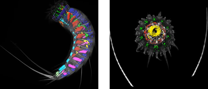
Researchers led by Dr. María Herranz from the Universidad Complutense de Madrid in Spain used Imaris to render the first complete three-dimensional reconstructions of microscopic marine invertebrates known as Kinorhynchs. These findings will enable scientists to better understand the evolution and diversity of body plans among animals.
Organisms in the phylum Kinorhyncha have a segmented body plan exhibiting cuticular structures along its length and a retractable introvert. These organisms are challenging to collect and work with in the laboratory and thus have not been well studied. Because of their relationship to other segmented animal groups such as arthropods, kinorhynchs can likely offer insight into how segmentation evolved.
The researchers studied the muscular anatomy, or myoanatomy, of Echinoderes, the most species-rich genus within Kinorhyncha. Comparing multiple species, which all exhibit a complex organ system in a segmented body, allowed the researchers to look for similarities and differences in muscle organization and determine whether variability among species of Echinoderes shows similar levels of variability within the phylum.
Taking a 3D view
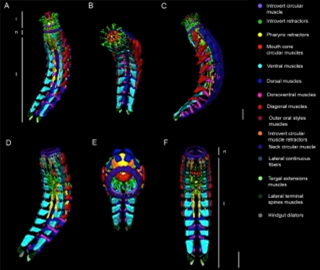
These 3D reconstructions show myoanatomy in Echinoderes horni. (A-C) Single specimen with the introvert fully everted. (D-F) Single specimen with the introvert fully retracted. Anterior to the top in all images. Abbreviations: i, introvert; n, neck; t, trunk. Scale bar, 30 μm. Reprinted from Herranz et al. Frontiers in Zoology 2014, 11:31.
“Imaris enabled us to assemble the confocal z-stacks in a straightforward intuitive way, with the potential to simultaneously view each of the separate components of an entire organ system in multiple dimensions (3D),” explains Dr. Herranz. “Imaris made it possible for us to count each muscle, describe their relative positions, detect their individual insertions along the body, and view a complete organ system integrated with other organs in the kinorhynch body. This would have been very difficult with 2D images.”
The researchers acquired confocal images of five echinoderid species and then used Imaris to create 3D reconstructions by surface-rendering the z-stacks. With Imaris, they digitally isolated and marked each group of muscles a specific color. This let them reconstruct the relationships of one muscle group to another and also build models of how particular myoanatomy most likely interacts during activities associated with feeding and motility of kinorhynchs within and between the sand grains where these tiny animals live.
Mapping muscle groups
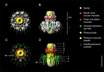
The head is one of the most complex regions of a kinorhynch and has multiple sets of overlapping muscles. The Imaris-created 3D maps of individual muscle groups within a larger functional unit let the researchers study the musculature of the head as well as the introvert. “As far as we know, this is the first comprehensive identification and interpretation of all the musculature present in this area,” said Dr. Herranz.
The 3D analyses showed that myoanatomy is highly conserved in Echinoderes. Among species, the researchers observed variation in the number and arrangement of introvert fibers and the composition of pharyngeal muscles. The researchers say that these detailed findings offer a model that can be used to understand the myoanatomy of closely related groups that have an introvert, such as priapulids, loriciferans, and some nematodes.
Research Paper: Herranz M, Boyle MJ, Pardos F, Neves RC. 2014. Comparative myoanatomy of Echinoderes (Kinorhyncha): a comprehensive investigation by CLSM and 3D reconstruction. Front Zool. 11(1):31. doi: 10.1186/1742-9994-11-31.